
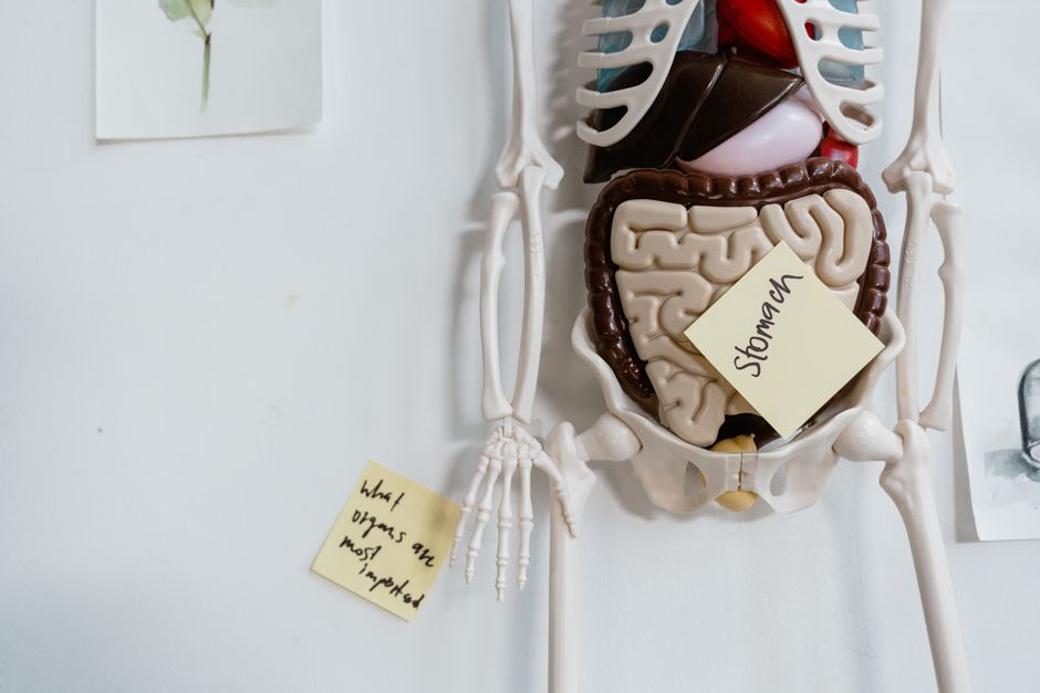
Your gut is not a smooth tube. On the inside, it looks more like a velvet carpet with millions of tiny finger-like projections called villi. These villi wave and move constantly, absorbing nutrients from food as it passes through. The surface is pink and moist, lined with a single layer of cells that act as a smart gatekeeper — deciding what gets into your bloodstream and what stays out.
What Does Your Gut Look Like On The Inside at a Microscopic Level?
Under a microscope, the inside of your gut is a complex landscape. The villi I mentioned are about 0.5 to 1.5 millimeters long. Each villus is covered in even smaller projections called microvilli. Together they create what scientists call the “brush border.”
This brush border increases the surface area of your small intestine by a factor of 30 to 60 times. Without it, your small intestine would be about the size of a studio apartment. With it, the surface area is roughly the size of a tennis court. That is where digestion and absorption actually happen.
Between the villi are deep pockets called crypts. These crypts produce new cells constantly. The cells lining your gut replace themselves every 3 to 5 days. That is the fastest cell turnover rate in your entire body. Your gut lining is essentially brand new every week.
What Does the Gut Lining Actually Look Like When Healthy?
A healthy gut lining looks plump and well-organized. The villi stand upright like tall grass in a healthy lawn. They are evenly spaced and densely packed. The cells themselves are tightly connected to each other with what are called tight junctions.
Think of tight junctions like zippers between cells. When they are working correctly, only small, fully digested nutrients can pass through. Larger food particles, bacteria, and toxins stay in the gut where they belong. The immune system below the lining stays calm because nothing foreign is getting through.
The color of a healthy gut lining is a rich pink. This comes from the dense network of blood vessels running just beneath the surface. These vessels carry away the nutrients your villi absorb. The mucus layer covering the lining is clear and slippery, protecting the cells from digestive enzymes and stomach acid.
| Feature | Healthy Gut | Unhealthy Gut |
|---|---|---|
| Villi shape | Tall, finger-like, upright | Short, blunted, flattened |
| Cell connections | Tight junctions sealed | Gaps between cells |
| Mucus layer | Thick, clear, protective | Thin, patchy, or absent |
| Color | Pink with visible blood flow | Pale or red and inflamed |
| Immune cells | Few, quiet | Many, active |
What Does an Unhealthy Gut Look Like on the Inside?
When the gut is damaged, the villi can become short and stubby. Instead of tall fingers reaching up to absorb nutrients, you see flattened nubs. This is called villous atrophy. Celiac disease is the most well-known cause of this. Research published in Gastroenterology has shown that untreated celiac disease can flatten villi almost completely.
The tight junctions between cells can also loosen. This is often called increased intestinal permeability, or “leaky gut” in popular media. The term is real in medical research, though its role in disease is still debated. What is clear is that when tight junctions open too wide, larger molecules pass through. The immune system below the lining sees these molecules as invaders and starts attacking.
The mucus layer can thin out too. A healthy mucus layer is about 150 micrometers thick in the small intestine and much thicker in the colon. When it gets thin, bacteria can touch the actual cells of the gut lining. That triggers inflammation. The CDC reports that chronic inflammation in the gut is linked to conditions like Crohn’s disease and ulcerative colitis.
Some people report that their gut feels “raw” or “burning” when they eat certain foods. While this is a real sensation, it does not always mean visible damage is present. Many people with gut symptoms have normal-looking villi and intact tight junctions on biopsy. Symptoms do not always match what the inside looks like.



What Causes the Gut to Change Appearance?
Several things can change how your gut looks on the inside. Diet is the most studied factor. A diet high in processed foods and low in fiber can reduce the diversity of gut bacteria. This bacterial community, called the microbiome, directly influences the health of your gut lining. The National Institutes of Health has funded research showing that fiber feeds the cells that produce mucus.
Infections are another cause. A bad case of food poisoning can temporarily damage villi. Most people recover within a week as new cells grow back. But repeated infections can cause lasting changes.
Medications matter too. Nonsteroidal anti-inflammatory drugs like ibuprofen and aspirin can damage the gut lining. Research in Alimentary Pharmacology & Therapeutics found that regular NSAID use can cause visible erosions and bleeding in the stomach and small intestine. These are not microscopic changes — doctors can see them during an endoscopy.
Alcohol is a direct irritant to the gut lining. It can thin the mucus layer and loosen tight junctions. Chronic heavy drinking is associated with visible changes in the gut lining that can last for months after stopping.
- Processed foods and low fiber reduce mucus production
- Infections can temporarily flatten villi
- NSAIDs cause visible erosions in the stomach and small intestine
- Alcohol thins the protective mucus layer
- Chronic stress alters blood flow to the gut lining
Can You See Your Own Gut on the Inside?
You cannot see your own gut without medical imaging. Some people try to use at-home tests that claim to show gut health. These tests analyze stool samples for bacteria, inflammation markers, or blood. They can provide useful information, but they do not show you what your gut looks like.
Doctors use two main methods to see the inside of your gut. An upper endoscopy uses a camera on a flexible tube passed through your mouth into your stomach and small intestine. A colonoscopy goes through the rectum to view the large intestine. During these procedures, doctors can take biopsies — tiny samples of tissue — to look at under a microscope.
Biopsy results are the gold standard for diagnosing conditions like celiac disease, Crohn’s disease, and microscopic colitis. No home test can replace that. If you are concerned about what your gut looks like on the inside, talk to a gastroenterologist. They can decide if a scope is appropriate based on your symptoms and medical history.
What Actually Improves the Appearance of Your Gut?
Evidence shows that dietary changes can improve the health and appearance of your gut lining. A gluten-free diet is the most dramatic example. In people with celiac disease, villi can regrow and return to normal height within 6 to 12 months of strict avoidance. This is well-documented in research from the American Journal of Gastroenterology.
For people without celiac disease, a diet rich in fermentable fiber helps. Foods like oats, bananas, onions, and garlic feed the bacteria that produce short-chain fatty acids. These fatty acids, especially butyrate, are the primary fuel source for the cells lining your colon. Higher butyrate levels are associated with a thicker mucus layer and tighter cell connections.
Some studies suggest that probiotics can support gut lining health. The evidence is moderate. A 2020 review in Nutrients found that certain strains of Lactobacillus and Bifidobacterium can strengthen tight junctions in animal studies. Human studies are less consistent. The strains matter, the dose matters, and individual response varies.
What does not have strong evidence? Bone broth, collagen supplements, and “gut healing” powders sold online. These are widely claimed to repair the gut lining, but as of 2026 there is no clinical evidence that they change the appearance of villi or tighten junctions in humans. Some people report feeling better after taking them, which is a real effect, but that does not mean the gut lining looks different.
Frequently Asked Questions
What does a healthy gut look like on the inside?
A healthy gut has tall, finger-like villi, a thick mucus layer, and tightly connected cells with no visible gaps or inflammation.
Can you see damage to your gut lining?
Damage is visible during an endoscopy or colonoscopy, and biopsies can confirm changes at the microscopic level.
How long does it take for gut villi to heal?
In celiac disease, villi can regrow within 6 to 12 months on a strict gluten-free diet, though recovery time varies by person.
Does leaky gut show up on a scope?
No, increased intestinal permeability is not visible during a scope and is measured through specialized lab tests rather than direct observation.
