
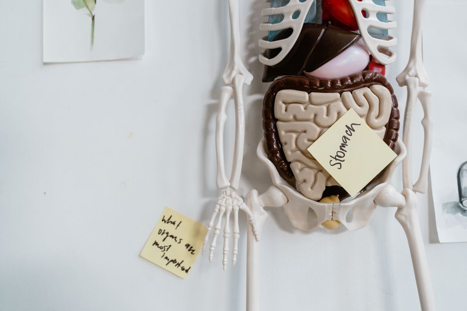
Your digestive system is a complex, coordinated tube made of four distinct tissue layers working together. From the inside out, these are the mucosa, submucosa, muscularis externa, and serosa. Each layer has a specific job, and together they move food, break it down, absorb nutrients, and push waste out of your body.
What Are the Four Main Tissue Layers of the Digestive Tract?
The wall of your digestive tract from your esophagus to your anus is built the same way. Think of it like a tube with four layers of pipe. Each layer is a different type of tissue with a different purpose.
The innermost layer is the mucosa. This is the layer that touches your food directly. It is a moist lining made of epithelial tissue. Its job is to secrete mucus, digestive enzymes, and hormones. It also absorbs nutrients once food is broken down. This layer is the first line of defense against anything harmful you swallow.
Next is the submucosa. This is a layer of connective tissue. It holds blood vessels, lymph vessels, and nerves. The submucosa supplies blood to the mucosa and carries away absorbed nutrients. It gives the digestive tract its stretch and strength.
The third layer is the muscularis externa. This is two layers of smooth muscle. One layer runs in circles around the tube. The other runs lengthwise. These muscles contract in waves, a process called peristalsis. Peristalsis pushes food and waste through your system. You do not control this muscle. It works automatically.
The outer layer is the serosa. This is a thin, slippery layer of connective tissue. It covers the outside of most digestive organs. It reduces friction so your stomach and intestines can slide against other organs as you move and digest food.
How Does the Mucosa Layer Actually Work?
The mucosa is where digestion and absorption happen. It is not just a simple lining. It has three parts of its own.
The surface is the epithelium. In your stomach, this epithelium secretes acid and digestive enzymes. In your small intestine, it is covered in tiny finger-like projections called villi. Each villus has even smaller projections called microvilli. These structures massively increase the surface area for absorbing nutrients. Research published in Gastroenterology has shown that the villi and microvilli increase the surface area of the small intestine by about 600 times.
Below the epithelium is the lamina propria. This is a thin layer of loose connective tissue. It holds small blood vessels and lymph vessels that pick up absorbed nutrients. It also contains immune cells. These cells act as a guard against bacteria and viruses that might slip through the epithelium.
The deepest part of the mucosa is the muscularis mucosae. This is a thin sheet of smooth muscle. It moves the mucosa itself. This movement helps mix food with digestive juices and brings the villi into contact with nutrients.
What Is the Role of the Submucosa and Its Nerves?
The submucosa is often overlooked, but it is critical. It is a tough layer of connective tissue that gives the digestive tract its ability to stretch and hold a meal.
Inside the submucosa is the submucosal plexus, also called Meissner’s plexus. This is a network of nerves. It is part of your enteric nervous system, sometimes called your “second brain.” This nerve network controls the function of the mucosa. It tells glands to secrete digestive juices and adjusts blood flow to the lining.
The submucosa also carries larger blood and lymph vessels. These vessels deliver oxygen to the digestive wall and carry away the nutrients that the mucosa absorbs. Without a healthy submucosa, the mucosa would starve and digestion would stop.


What Tissues Make Up Your Digestive System’s Muscle Layers?
The muscularis externa is the layer that physically moves your food. It is made of smooth muscle tissue, which is different from the muscle in your arms or legs. Smooth muscle works automatically and does not tire easily.
This layer has two sub-layers. The inner layer has muscle fibers running in circles around the tube. When these contract, they squeeze the tube shut. The outer layer has fibers running lengthwise. When these contract, they shorten the tube.
Between these two muscle layers is the myenteric plexus, or Auerbach’s plexus. This is another nerve network. It coordinates the contractions of the two muscle layers. It is the main driver of peristalsis. The myenteric plexus is so important that damage to it from diabetes or surgery can cause serious motility problems.
The stomach has a third layer of muscle in the muscularis externa. This extra layer runs at an angle and helps the stomach churn and mix food into a paste called chyme.
How Does the Serosa Protect Your Digestive Organs?
The serosa is the outermost layer. It is a single layer of flat cells called mesothelium sitting on a thin layer of connective tissue. It is smooth and slippery.
The serosa secretes a thin, watery fluid. This fluid acts like a lubricant. It allows your stomach and intestines to move and slide against each other and against the walls of your abdominal cavity. Without this lubrication, every movement would cause friction and pain.
In some parts of the digestive system, the serosa is replaced by a layer called the adventitia. This happens in the esophagus and the rectum. The adventitia is just connective tissue that anchors these organs to nearby structures. It is less slippery because these parts do not need to move as freely.
| Layer | Main Tissue Type | Primary Function |
|——-|——————|——————|
| Mucosa | Epithelium, connective, smooth muscle | Secretion, absorption, protection |
| Submucosa | Connective tissue, nerves, blood vessels | Support, blood supply, nerve signals |
| Muscularis externa | Smooth muscle (2-3 layers) | Peristalsis, mixing, movement |
| Serosa | Mesothelium, connective tissue | Lubrication, friction reduction |
What About the Organs That Are Not Part of the Tube?
Your digestive system includes more than just the long tube from mouth to anus. Several solid organs help digestion but are not part of the tube wall itself. These organs are made of different tissues.
The liver is the largest internal organ. It is made mostly of hepatocytes, which are specialized liver cells. The liver produces bile, which helps break down fats. It also processes nutrients that the intestines absorb.
The pancreas is made of exocrine and endocrine tissue. The exocrine part produces digestive enzymes that break down proteins, fats, and carbohydrates. The endocrine part produces hormones like insulin that control blood sugar.
The gallbladder is a small sac made of smooth muscle and epithelial tissue. It stores and concentrates bile from the liver. When you eat a fatty meal, it contracts and releases bile into the small intestine.
These organs connect to the main digestive tube through ducts. They are not made of the same four layers as the tube. But they are essential for complete digestion.
Frequently Asked Questions
Frequently Asked Questions
Which tissue layer is responsible for peristalsis?
The muscularis externa is the layer responsible for peristalsis. Its two layers of smooth muscle contract in coordinated waves to push food forward.
What is the difference between the mucosa and the submucosa?
The mucosa is the innermost lining that secretes enzymes and absorbs nutrients. The submucosa is the connective tissue layer underneath that supplies blood and nerves to the mucosa.
Does the serosa cover all digestive organs?
No. The serosa covers most digestive organs inside the abdominal cavity. The esophagus and rectum have an adventitia instead, which anchors them to surrounding tissues.
Can the enteric nervous system work without the brain?
Yes, to a large degree. The enteric nervous system in the submucosa and muscularis can coordinate digestion independently. The brain does influence it, but the gut can function on its own.
