
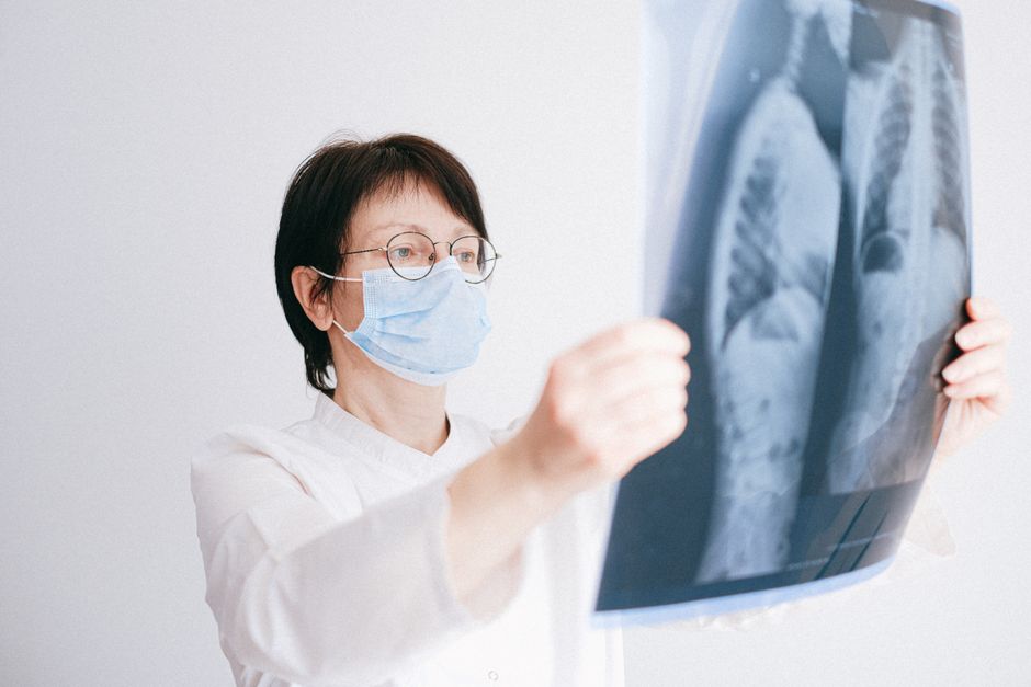
Testing for Aspergillus in the lungs is not a single test. It usually requires a combination of imaging, blood tests, and lab analysis of lung fluid or tissue. The right method depends on the type of Aspergillus condition suspected, which includes allergic forms, chronic infections, and invasive disease in people with weakened immune systems.
What Is Aspergillus and Why Does Testing Matter?
Aspergillus is a common mold found indoors and outdoors. Most people breathe in Aspergillus spores every day without getting sick. The immune system clears them quickly.
Problems arise in specific groups. People with asthma or cystic fibrosis may develop allergic reactions. People with damaged lungs from prior disease may develop chronic infections. People with severely weakened immune systems — such as organ transplant recipients or those on chemotherapy — face the highest risk of invasive aspergillosis, where the fungus spreads through lung tissue and beyond.
Because the symptoms overlap with other lung conditions, testing must be precise. A chest X-ray alone cannot confirm Aspergillus. Neither can a physical exam. The diagnostic process is layered and deliberate.
How To Test For Aspergillus In The Lungs Key Methods: Imaging First
A chest CT scan is typically the first imaging step when a doctor suspects Aspergillus. A standard chest X-ray often misses early changes in the lungs.
CT scans reveal specific patterns. In invasive aspergillosis, a “halo sign” — a dark ring around a lung nodule — may appear in early stages. In chronic forms, cavities and thickening of lung tissue are common. In allergic bronchopulmonary aspergillosis (ABPA), the scan may show mucus plugs or airways that are wider than normal.
Imaging alone is never enough for a diagnosis. But it guides the next steps. A doctor may see something suspicious and order blood tests or a bronchoscopy based on what the CT shows.
Blood Tests for Aspergillus Antibodies and Antigens
Blood tests detect the immune system’s response to the fungus or the fungus itself. Several different tests exist, and each one serves a different purpose.
Aspergillus IgG antibodies measure past exposure. High levels suggest the immune system has met the fungus before. This test is most useful for chronic pulmonary aspergillosis, where the body has been fighting the fungus over a long period.
Aspergillus IgE antibodies measure allergic response. Elevated levels point to allergic conditions like ABPA. This test is often paired with a total IgE measurement, which is also elevated in ABPA.
Galactomannan antigen testing detects a sugar molecule on the surface of the Aspergillus fungus. This test is particularly useful in invasive disease, especially in patients with weakened immune systems. It can be done on blood or on fluid from a lung wash. The test is less helpful in allergic or chronic forms because the fungus is not actively growing in the bloodstream in those conditions.
Blood tests are non-invasive and relatively quick. But they do not always give a clear answer. False negatives happen, especially early in infection. False positives can occur in people who have received certain antibiotics or who have other fungal infections.





Sputum Culture and Microscopy
A sputum sample — mucus coughed up from the lungs — can be examined under a microscope and cultured in a lab. If Aspergillus grows from the sample, that is strong evidence the fungus is present in the airways.
However, a positive culture does not mean active disease. Aspergillus can colonize the airways of people with chronic lung disease without causing infection. The culture result must be interpreted alongside symptoms, imaging, and blood tests.
Also, sputum samples miss the fungus some of the time. The organism may be present in the lungs but not in the sample coughed up. For this reason, a negative sputum culture does not rule out Aspergillus.
In some cases, a doctor may request a bronchoscopy to obtain a better sample. During this procedure, a thin tube with a camera is passed through the nose or mouth into the airways. Saline is introduced and then suctioned back out, collecting fluid from deeper in the lungs. This is called a bronchoalveolar lavage, or BAL. The fluid can be cultured, examined under a microscope, and tested for galactomannan.
When Is a Lung Biopsy Needed?
A lung biopsy is the most definitive test for invasive aspergillosis. A small piece of lung tissue is removed and examined under a microscope. If fungal filaments — called hyphae — are seen growing into lung tissue, the diagnosis is confirmed.
Biopsies are invasive. They carry risks including bleeding and collapsed lung. For this reason, they are reserved for cases where the diagnosis remains uncertain after less invasive testing, or when the clinical picture demands certainty.
In practice, many doctors treat invasive aspergillosis based on a combination of risk factors, imaging, and blood tests without a biopsy. This is common because patients with invasive disease are often too ill for invasive procedures. But when a biopsy is performed and shows the fungus invading tissue, that is the gold standard.
PCR Testing for Aspergillus DNA
Polymerase chain reaction, or PCR, detects Aspergillus genetic material in blood, sputum, or lung fluid. The test is highly sensitive — it can find very small amounts of fungal DNA.
PCR is particularly valuable in invasive aspergillosis, where early detection improves outcomes. It is also used to monitor response to treatment in some cases.
One challenge with PCR is that it can detect DNA from dead fungus or from spores that were simply inhaled and not causing disease. A positive PCR result must be considered in context. It is a helpful tool, but not a standalone diagnosis.
Some research suggests combining PCR with galactomannan testing improves diagnostic accuracy. Each test catches different aspects of the infection, and together they provide a fuller picture.
Comparing the Main Testing Methods
| Test | What It Detects | Best Used For | Limitations |
|---|---|---|---|
| Chest CT scan | Structural changes in lung tissue | Initial evaluation; guides further testing | Cannot confirm the fungus is the cause |
| Aspergillus IgG | Past exposure antibodies | Chronic pulmonary aspergillosis | Not useful in early or invasive disease |
| Aspergillus IgE | Allergic response antibodies | ABPA and allergic conditions | Does not detect infection |
| Galactomannan antigen | Fungal cell surface molecule | Invasive aspergillosis | False negatives early; false positives with some antibiotics |
| Sputum culture | Live fungus growth | Confirming fungal presence in airways | Positive result may mean colonization, not disease |
| PCR | Fungal DNA | Invasive disease; monitoring treatment | Detects dead fungus; cannot distinguish colonization from infection |
| Lung biopsy | Fungal invasion into tissue | Definitive diagnosis | Invasive; carries bleeding and collapse risk |
How Doctors Combine Test Results
No single test is perfect. Doctors combine multiple pieces of evidence to reach a diagnosis.
For allergic forms like ABPA, the diagnosis rests on symptoms, elevated IgE levels, positive Aspergillus antibodies, and imaging findings. For chronic pulmonary aspergillosis, the picture includes long-term symptoms, cavities on CT, and positive IgG antibodies. For invasive disease, the combination typically involves risk factors, characteristic CT findings, and a positive galactomannan or PCR result.
Clinical context matters more than any individual test result. A positive blood test in a person with no symptoms and a normal CT scan usually means nothing. A negative blood test in a person with classic symptoms and typical imaging does not rule out the disease.
Testing for Aspergillus is a process of building a case. Each result adds information, and the doctor weighs all of it together.
What to Expect During Testing
Blood tests are simple and quick — just a standard blood draw. Sputum collection requires coughing deeply to bring up mucus from the lungs. This can take several attempts.
Bronchoscopy is done under sedation. The procedure typically takes 30 to 60 minutes, and most people go home the same day. Sore throat and mild coughing are common afterward.
Lung biopsy is a more involved procedure. It may be done through the chest wall with a needle, guided by CT imaging, or during a bronchoscopy. Recovery time varies depending on the approach.
Results take different amounts of time. Blood antibody tests may return within a few days. Cultures need time for the fungus to grow — often one to two weeks. PCR results can be available in one to two days. Biopsy results depend on how the tissue is processed, generally several days.
Frequently Asked Questions
Can a chest X-ray detect Aspergillus in the lungs?
A chest X-ray can show abnormalities but cannot confirm Aspergillus. A CT scan provides much more detail and is the preferred imaging method.
How long does it take to get Aspergillus test results?
Blood tests typically take a few days, while fungal cultures can take one to two weeks. PCR results may be available within one to two days.
Is a lung biopsy always required to diagnose Aspergillus?
No. Many cases are diagnosed using imaging, blood tests, and fluid samples. Biopsy is reserved for uncertain cases or when a definitive answer is needed.
Can Aspergillus testing give false results?
Yes. False negatives occur early in infection, and false positives can happen with certain antibiotics or when the fungus is present but not causing disease.
