
Yes, a CT scan can detect gallstones, but it is not the best test for finding them. Many gallstones contain enough calcium to show up clearly on a CT scan. However, some gallstones are made mostly of cholesterol and can be nearly invisible on a CT scan. This is why doctors often prefer an ultrasound when they suspect gallstones.
How Does a CT Scan Detect Gallstones?
A CT scan uses X-rays to create detailed cross-section images of your body. It is excellent at showing bones, blood vessels, and solid organs like the liver and pancreas. When it comes to the gallbladder, the results depend on what the gallstones are made of.
Gallstones form when bile hardens in the gallbladder. Bile is a digestive fluid made by the liver. There are two main types of gallstones:
- Cholesterol stones — These are the most common type. They are usually yellow-green and contain very little calcium. On a CT scan, they can look like the surrounding bile, making them hard to spot.
- Pigment stones — These are darker and smaller. They form when there is too much bilirubin in the bile. They often contain enough calcium to be seen on a CT scan.
Studies have shown that CT scans miss a significant number of gallstones, especially the cholesterol type. Some research suggests CT scans detect only about 50-75% of gallstones. This means a CT scan that looks normal does not rule out gallstones.
Why Is Ultrasound the Preferred Test?
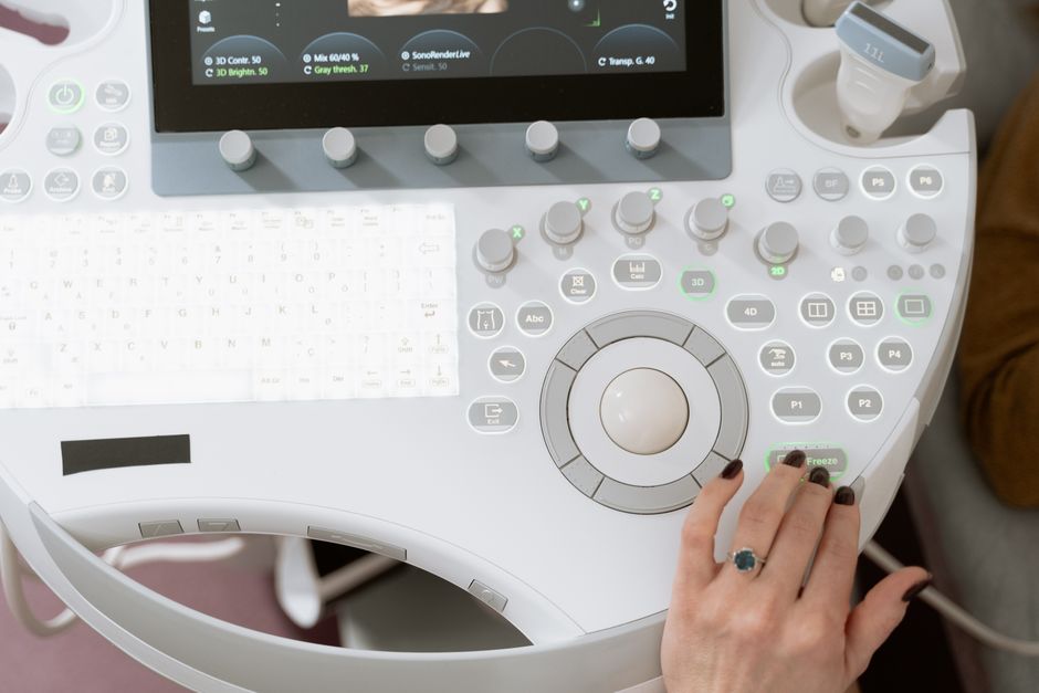
Abdominal ultrasound is the standard first test for gallstones. It uses sound waves to create images of your organs in real time. Ultrasound is highly accurate for detecting gallstones — even small ones. It can also show signs of gallbladder inflammation, like a thickened gallbladder wall or fluid around the organ.
Ultrasound has several advantages over CT for this specific problem:
- It detects gallstones with greater accuracy, often above 95%.
- It does not expose you to radiation.
- It is faster and less expensive.
- It can be done at the bedside if you are very ill.
CT scans are generally used when the diagnosis is unclear or when doctors suspect complications. For example, if you have severe abdominal pain and the ultrasound is normal, a CT scan can help look for other causes like appendicitis, diverticulitis, or a bowel blockage.
When Is a CT Scan Actually Useful for Gallbladder Problems?
CT scans play an important role in gallbladder disease, but not as the first test. They are most valuable when doctors need to look beyond the gallbladder itself. A CT scan can show complications that ultrasound might miss.
These complications include:
- Perforation — A hole in the gallbladder wall.
- Abscess — A pocket of infection near the gallbladder.
- Gallstone ileus — A rare condition where a large stone blocks the intestine.
- Pancreatitis — Inflammation of the pancreas, which can be caused by a stone blocking the common bile duct.
A CT scan is also helpful when the ultrasound images are unclear. This can happen if you have a lot of bowel gas or if you are overweight. In these situations, the CT scan provides a more complete view of the abdomen.





Can You See Gallstones On A Ct Scan of the Abdomen and Pelvis?
Gallstones are located in the upper right part of the abdomen. A CT scan of the abdomen will include the gallbladder. A CT scan of the pelvis only looks at the lower part of your body and will not show the gallbladder.
If your doctor orders a CT scan of the abdomen for an unrelated reason — like a kidney stone check — the radiologist may notice gallstones as an incidental finding. This happens fairly often. Many people have gallstones without any symptoms. In these cases, no treatment is needed unless the stones cause problems.
It is important to note that a CT scan is not the right tool to check the bile ducts. The bile ducts are small tubes that carry bile from the liver and gallbladder to the intestine. Stones in the bile ducts can cause jaundice and serious infections. An MRI called an MRCP is the best imaging test for bile duct stones. An endoscopic ultrasound is another accurate option.
What Does a Gallstone Look Like on a CT Scan?
Gallstones appear differently on CT scans depending on their composition. Radiologists describe them based on their density, which shows up as different shades of gray on the images.
Calcified stones appear bright white on a CT scan. They are easy to see. Cholesterol stones appear dark or similar to the bile around them. They can be missed entirely. Some stones have a mixed appearance with layers of calcium and cholesterol.
Sometimes a CT scan shows indirect signs of gallstones even when the stones themselves are not visible. These signs include a swollen gallbladder, thickened walls, or air in the gallbladder wall. These findings suggest inflammation and prompt further testing.
Because of these limitations, a negative CT scan never completely rules out gallbladder disease. If your symptoms strongly suggest gallstones but the CT scan is normal, your doctor will likely order an ultrasound or an MRCP.
What Are the Symptoms of Gallstones?
Many people with gallstones never have symptoms. These are called silent gallstones. They are usually found by accident during imaging for another reason and typically require no treatment.
Symptomatic gallstones cause a specific type of pain called biliary colic. This pain is usually:
- Located in the upper right abdomen or the center of the upper belly
- Severe and steady
- Worse after eating a fatty meal
- Often felt in the back or right shoulder blade
- Lasts from 30 minutes to several hours
You should seek emergency care if you have pain lasting more than five hours, fever, chills, yellowing of the skin or eyes, or vomiting. These can be signs of acute cholecystitis — an inflamed gallbladder — or a blocked bile duct. Both conditions require prompt medical treatment.
How Are Gallstones Treated?
Treatment depends on whether you have symptoms. Silent gallstones usually need no treatment. Your doctor will simply monitor them.
Symptomatic gallstones are typically treated with surgery to remove the gallbladder. This operation is called a cholecystectomy. It is one of the most common surgeries performed in the United States. Most are done laparoscopically, which means small incisions and a shorter recovery time.
Removing the gallbladder does not affect your ability to digest food in a major way. The gallbladder stores bile, but your liver continues to make it. Bile flows directly from the liver into the intestine after surgery. Some people experience loose stools after eating fatty meals, but most adapt within a few months.
Medications to dissolve gallstones exist, but they are rarely used. They work only for small cholesterol stones and can take months or years to work. Stones often return after stopping the medication. Surgery remains the most effective and definitive treatment for symptomatic gallstones.
What Happens If Gallstones Are Left Untreated?
Untreated gallstones can lead to serious complications. The most common is acute cholecystitis, an infection and inflammation of the gallbladder. This condition requires antibiotics and usually surgery. Without treatment, the gallbladder can rupture, which is a medical emergency.
Gallstones can also travel into the bile ducts and block the flow of bile. This causes jaundice — yellowing of the skin and eyes — and can lead to a dangerous infection called cholangitis. A blocked pancreatic duct can cause pancreatitis, a painful and potentially life-threatening inflammation of the pancreas.
These complications are why doctors take gallstone symptoms seriously. If you have recurrent pain in the upper right abdomen, especially after meals, talk to your doctor. Early diagnosis with ultrasound and prompt treatment can prevent most serious outcomes.
Frequently Asked Questions
Can a CT scan miss gallstones?
Yes. CT scans miss many gallstones, especially cholesterol stones that lack calcium. Ultrasound is significantly more accurate for detecting gallstones.
What is the best scan to detect gallstones?
An abdominal ultrasound is the best first test for gallstones. It is highly accurate, safe, and widely available. An MRCP is the best test for stones in the bile ducts.
Do gallstones show up on CT without contrast?
Some do, but not all. Calcified stones are visible on a non-contrast CT scan. Cholesterol stones may be invisible. Contrast is not typically needed to see gallstones themselves.
Can a CT scan show gallbladder inflammation?
Yes. A CT scan can show a thickened gallbladder wall, fluid around the gallbladder, and other signs of inflammation. However, ultrasound remains the preferred test for diagnosing acute cholecystitis.
