
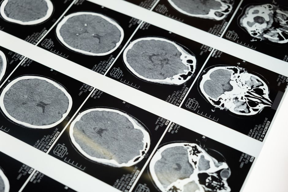
A T2 weighted MRI scan is a specific type of magnetic resonance imaging that highlights water content in your body’s tissues. It creates images where areas with more water appear bright, while denser tissues like bone appear dark. This makes it an excellent tool for spotting inflammation, tumors, infections, and injuries that other scans might miss.
Think of it like a map where water glows. Doctors use T2 weighted scans to see swelling, fluid-filled cysts, and areas of disease activity. The “weighted” part means the machine is tuned to emphasize how water molecules behave in a magnetic field. It is not a separate machine from a standard MRI, just a different setting.
If you have ever had an MRI of your knee after an injury or a brain scan for headaches, you likely had a T2 weighted sequence done. It is one of the most common and useful MRI techniques in medicine today.
How Is a T2 Weighted MRI Different From a T1 Weighted MRI?
T1 and T2 weighted scans are two sides of the same coin. They both use the same MRI machine but measure different physical properties of tissue. The main difference is what shows up bright and what shows up dark.
On a T1 weighted scan, fat appears bright and water appears dark. This makes it great for looking at anatomy, like the structure of organs or the outline of a tumor. On a T2 weighted scan, the opposite is true: water appears bright and fat appears dark.
Here is a simple comparison table to make the difference clear:
| Tissue Type | T1 Weighted Appearance | T2 Weighted Appearance |
|---|---|---|
| Water (CSF, urine) | Dark | Bright |
| Fat | Bright | Dark |
| Muscle | Gray | Gray |
| Inflammation/Edema | Dark | Bright |
| Bone | Dark | Dark |
Doctors often order both T1 and T2 sequences during the same MRI exam. The T1 gives a clear picture of anatomy, and the T2 highlights any abnormal fluid or swelling. Together, they provide a complete picture that neither could give alone.
What Does a T2 Weighted MRI Scan Show in the Brain?
In brain imaging, T2 weighted scans are invaluable. They show areas of the brain where water has accumulated, which often signals disease or injury. Multiple sclerosis lesions, for example, appear as bright white spots on T2 images because they contain inflammation and fluid.
Research published in the journal Radiology has shown that T2 weighted imaging detects over 90% of MS lesions visible on MRI. This is why it is a standard part of diagnosing and monitoring the disease. Strokes also show up well on T2 scans, though they may take several hours to become visible.
Brain tumors, infections like abscesses, and areas of swelling from trauma all appear bright on T2. The scan does not tell the doctor exactly what the bright spot is, but it tells them something is wrong and where to look further. This is why T2 is often called a “screening” sequence.
One thing many people do not realize is that normal aging also causes some bright spots on T2 brain scans. These are called white matter hyperintensities and are common in people over 60. They do not always mean disease, but they can be linked to high blood pressure or small vessel disease.
What Does a T2 Weighted MRI Show in the Spine and Joints?
In the spine, T2 weighted scans are the gold standard for looking at discs. A healthy disc contains a lot of water and appears bright. A degenerated or herniated disc loses water and appears darker. This makes it easy for radiologists to spot disc problems.
Spinal cord compression, inflammation, and tumors also show up clearly. If you have back pain with nerve symptoms like numbness or weakness, a T2 weighted MRI is often the first imaging test ordered. The bright fluid around the spinal cord also helps doctors see if there is a blockage or infection.
In joints like the knee or shoulder, T2 scans highlight fluid in places it should not be. A torn meniscus in the knee, for example, may show a bright line of fluid extending into the dark cartilage. Ligament tears, like an ACL injury, also show up because of the fluid around the torn fibers.
Arthritis causes joint swelling, which appears bright on T2. The scan can show inflammation in the lining of the joint, called synovitis, even before it causes bone damage. This allows for earlier treatment in conditions like rheumatoid arthritis.

What Are the Risks and Limitations of T2 Weighted MRI?
T2 weighted MRI scans are very safe for most people. They use no ionizing radiation, unlike X-rays or CT scans. The magnetic field and radio waves have no known long-term health risks. The main safety concerns come from metal in the body.
People with certain implanted devices cannot have an MRI. This includes older pacemakers, cochlear implants, and some aneurysm clips. The strong magnetic field can move or heat these objects. Always tell your doctor about any metal in your body before an MRI.
Another limitation is that T2 scans are not specific. A bright spot on a T2 image could be inflammation, a tumor, an infection, or scar tissue. The radiologist needs other sequences or clinical information to narrow it down. This is why you rarely get just a T2 scan alone.
Some people feel claustrophobic inside the machine. The scan also requires you to lie very still for 20 to 45 minutes. Movement can blur the images and require a repeat scan. Open MRI machines exist but often produce lower quality T2 images.
Contrast agents like gadolinium are sometimes used with T2 scans to improve detail. The FDA has warned that gadolinium can remain in the body, though no definite harm has been proven from this retention. Discuss with your doctor whether contrast is truly needed for your case.
Does What Is A T2 Weighted MRI Scan and What Does It Show Work Better Than Other Scans?
No single scan is best for everything. T2 weighted MRI excels at showing soft tissue and fluid, making it ideal for inflammation, tumors, and injuries. But it is not the best choice for every situation.
For bone fractures, a CT scan is faster and more detailed. For lung problems, a chest X-ray or CT is better because MRI does not image air well. For acute bleeding in the brain, a CT scan is usually the first choice because it is faster and more sensitive to fresh blood.
Research from the American College of Radiology guidelines shows that T2 weighted MRI is the preferred test for many conditions including:
- Multiple sclerosis diagnosis and monitoring
- Spinal cord compression
- Knee meniscal tears
- Brain tumors (with contrast)
- Inflammatory arthritis
- Prostate cancer detection
T2 scans are also very good at ruling things out. A normal T2 scan of the brain, for example, makes a large tumor or active MS highly unlikely. This “rule-out” power is one reason doctors order them so frequently.
The bottom line is that T2 weighted MRI is not better than all other scans, but it is the best tool for a specific set of problems. Your doctor chooses the right scan based on what they suspect is wrong.
How Should You Prepare for a T2 Weighted MRI Scan?
Preparation for a T2 weighted MRI is the same as for any MRI. You do not need to fast or change your diet unless your doctor specifically says so. The main preparation is removing all metal objects from your body.
Before the scan, you will fill out a safety questionnaire. Be honest about any surgeries, implants, or metal in your body. This includes dental work, piercings, and tattoos. Some older tattoo inks contain iron, which can cause skin irritation during the scan.
Wear loose, comfortable clothing without metal zippers or buttons. You may be given a hospital gown to wear. Remove all jewelry, watches, and credit cards, as the magnetic field can damage them. Hearing aids and dentures also need to come out.
If you are claustrophobic, talk to your doctor beforehand. They may prescribe a mild sedative or offer an open MRI if available. You can also ask for earplugs or headphones, as the machine makes loud knocking noises during the scan.
Tell the technician if you are pregnant or could be pregnant. MRI is generally considered safe during pregnancy, but it is usually avoided in the first trimester unless absolutely necessary. The technician will walk you through every step and you can communicate with them during the scan.
Frequently Asked Questions
How long does a T2 weighted MRI scan take?
A T2 weighted sequence itself takes about 3 to 5 minutes. The full MRI exam, which includes multiple sequences, typically takes 20 to 45 minutes total.
Can I eat before a T2 weighted MRI?
Yes, you can eat and drink normally unless your doctor tells you otherwise. Some scans of the abdomen or pelvis may require fasting, but this is not related to the T2 sequence.
Is a T2 weighted MRI painful?
No, the scan itself is painless. You may feel uncomfortable lying still, and some people find the machine noise annoying. If you have claustrophobia, tell your doctor in advance.
What does a bright spot on a T2 MRI mean?
A bright spot means there is more water in that area than normal. This could be inflammation, swelling, a tumor, infection, or scar tissue. Your radiologist and doctor will interpret the finding based on your symptoms and other images.
