
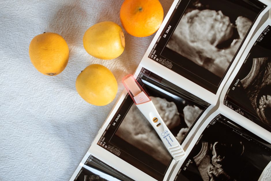
A choroid plexus ultrasound is a specialized imaging test that uses high-frequency sound waves to examine the choroid plexus — the network of blood vessels inside the brain’s fluid-filled spaces. The test is most commonly done during pregnancy as part of a routine fetal anatomy scan, but it is also used after birth in newborns and, less often, in adults. It is painless, uses no radiation, and typically takes only a few minutes once the scan begins.
What Is the Choroid Plexus and Why Does It Matter?
The choroid plexus is a small, vascular structure located inside the brain’s ventricles — the fluid-filled cavities that hold cerebrospinal fluid, or CSF. It produces most of the CSF that surrounds and cushions the brain and spinal cord.
Because the choroid plexus sits inside fluid, it shows up clearly on ultrasound. That makes it a useful landmark for anyone examining the brain with sound waves, whether in a fetus, a newborn, or an adult.
On ultrasound, the choroid plexus usually appears as a bright, echogenic structure. Echogenic means it reflects sound waves strongly and shows up lighter than surrounding tissue. This bright appearance is normal and expected.
The choroid plexus is also where cysts sometimes form. Most of these cysts are benign and resolve on their own. Its visibility and its link to certain findings are why it gets specific attention during imaging.
What To Expect From a Choroid Plexus Ultrasound During Pregnancy
During pregnancy, the choroid plexus is examined as part of the standard fetal anatomy survey, usually performed in the second trimester. The sonographer does not usually perform a separate dedicated scan just for the choroid plexus unless something specific prompts it.
Here is what the experience typically involves:
- You lie on an exam table, often on your back or slightly tilted to one side.
- A gel is applied to your abdomen to help transmit sound waves.
- A transducer is moved across your belly to capture images.
- The scan is painless. You may feel mild pressure from the probe.
- Results are usually reviewed by a radiologist or maternal-fetal medicine specialist.
The scan itself usually takes 20 to 45 minutes for a full anatomy survey. The choroid plexus portion is only a small part of that time.
If the sonographer focuses specifically on the choroid plexus, it is often because a choroid plexus cyst was seen or is being followed. These cysts appear as small, fluid-filled spaces within the choroid plexus.
What Does a Choroid Plexus Cyst Found on Ultrasound Mean?
A choroid plexus cyst is a small fluid-filled pocket inside the choroid plexus. It is one of the most common findings on a second-trimester ultrasound.
In most cases, an isolated choroid plexus cyst — meaning one found without any other abnormal findings — is not associated with any health problem. The cyst typically disappears on its own before or shortly after birth. It does not affect brain development or function.
The picture changes when a choroid plexus cyst appears alongside other findings. In that situation, clinicians may recommend additional testing, because certain chromosomal conditions have been associated with choroid plexus cysts when other markers are also present.
This is why context matters so much. A cyst on its own and a cyst with other findings are two very different situations. The ultrasound report will usually note whether the cyst is isolated or whether other features were seen.
Some clinicians recommend follow-up ultrasound to confirm the cyst has resolved. Others consider an isolated cyst a normal variant that needs no further imaging. Practice varies, and both approaches exist in current clinical care.




How Is a Choroid Plexus Ultrasound Done in a Newborn?
In newborns, a choroid plexus ultrasound is usually performed through the anterior fontanelle — the soft spot on the top of the baby’s head that has not yet closed into solid bone. This opening gives sound waves a clear path into the brain.
This type of scan is called a cranial ultrasound. It is commonly used in premature infants and in babies who have had a difficult delivery or show concerning neurological signs. It can detect bleeding in the brain, fluid buildup, and structural abnormalities.
The procedure is gentle. The baby usually lies on their back while a small probe is moved over the soft spot. Most scans take 10 to 20 minutes. No sedation is typically needed.
Because the fontanelle closes as an infant grows, this window for imaging is limited. After the soft spot closes, usually by around 12 to 18 months, ultrasound can no longer easily reach the brain, and other imaging methods are used instead.
Can Adults Have a Choroid Plexus Ultrasound?
In adults, ultrasound cannot penetrate the skull bone well enough to image the brain clearly. For that reason, a choroid plexus ultrasound is rarely used in adults.
When adults need imaging of the choroid plexus, doctors typically use MRI or CT instead. MRI in particular can show the choroid plexus in detail and is the preferred method for evaluating most brain structures in adults.
There is one exception worth noting. During brain surgery, surgeons sometimes use intraoperative ultrasound directly on the brain surface. This is a specialized use and not the same as a standard diagnostic scan.
What Are the Risks and Limitations?
Diagnostic ultrasound is considered safe when used appropriately. It does not use ionizing radiation, which distinguishes it from CT scans and X-rays. No consistent evidence links diagnostic ultrasound during pregnancy to harm to the fetus.
That said, ultrasound has real limitations:
- It cannot see through bone, which limits its use in older children and adults.
- Image quality depends on the equipment, the operator, and the position of the fetus or patient.
- Some findings, like choroid plexus cysts, require context from other tests to interpret correctly.
- Ultrasound cannot rule out every possible condition.
Ultrasound is generally not used for prolonged or repeated scanning without a medical reason. The principle of keeping exposure as low as reasonably achievable applies, even though no harm has been demonstrated at diagnostic levels.
How to Prepare and What Results Mean
Preparation for a fetal choroid plexus ultrasound is minimal. You may be asked to drink water and avoid urinating before the scan, depending on the clinic’s protocol. Loose, comfortable clothing is helpful.
For a newborn cranial ultrasound, no special preparation is usually needed. Feeding the baby shortly before the scan can help them stay calm.
Results are interpreted by a radiologist or specialist. The report will describe what was seen and whether any follow-up is recommended. If a choroid plexus cyst is found, the report should note whether it is isolated or accompanied by other findings.
If you receive a report mentioning a choroid plexus cyst, ask your clinician these questions:
- Was the cyst isolated, or were other findings present?
- Is any additional testing recommended?
- Is follow-up imaging planned, and if so, when?
Understanding the context of any finding is more useful than focusing on the finding alone. A single ultrasound result rarely tells the whole story.
Frequently Asked Questions
Is a choroid plexus ultrasound painful?
No, it is not painful. You or your baby may feel mild pressure from the ultrasound probe, but there are no needles, incisions, or injections involved.
Does a choroid plexus cyst mean something is wrong with my baby?
In most cases, an isolated choroid plexus cyst is not linked to any health problem and resolves on its own. When other findings are also present, additional testing may be recommended to assess the full picture.
How long does a choroid plexus ultrasound take?
The choroid plexus portion of a scan takes only a few minutes. A full fetal anatomy survey usually takes 20 to 45 minutes, while a newborn cranial ultrasound typically takes 10 to 20 minutes.
Can a choroid plexus ultrasound detect Down syndrome?
No, ultrasound alone cannot diagnose Down syndrome or other chromosomal conditions. A choroid plexus cyst may be one marker considered alongside other findings, but definitive diagnosis requires genetic testing such as amniocentesis or cell-free DNA screening.
