
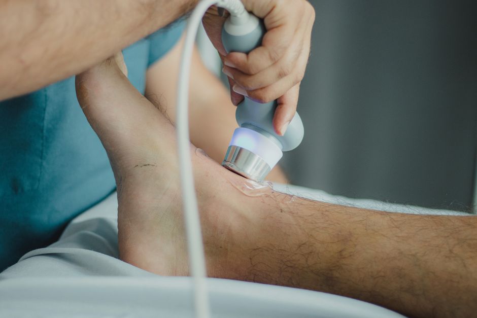
An OB ultrasound (obstetric ultrasound) is a medical imaging test that uses sound waves to create pictures of a developing baby inside the womb. It is a routine part of prenatal care. There are two main types: transabdominal (over the belly) and transvaginal (inside the vagina). Obstetric ultrasound is used to confirm pregnancy, check the baby’s growth, find the due date, look for birth defects, and monitor the placenta and amniotic fluid. During the scan, you lie on a table while a handheld device glides over your belly. The procedure is painless and there is no radiation. Expect to have a full bladder for early scans and to wear loose clothing. The whole visit usually takes 20 to 45 minutes.
What Is an OB Ultrasound Types Uses What to Expect
An obstetric ultrasound is a safe and common imaging tool used throughout pregnancy. It works by sending high-frequency sound waves into the body. Those waves bounce off tissues and the baby, and a computer turns the echoes into live images on a screen. There is no ionizing radiation involved, which makes it safe for both mother and baby. The test is performed by a trained sonographer or a doctor. It is also called a sonogram or prenatal ultrasound.
There are two main types of OB ultrasound. The type your provider uses depends on how far along the pregnancy is and what information they need.
What Are the Different Types of OB Ultrasound?
Transabdominal ultrasound is the most common type. The sonographer applies a water‑based gel to your lower belly and glides a handheld transducer over the skin. This type gives a broad view of the uterus and baby. It works best after about 10 to 12 weeks of pregnancy.
Transvaginal ultrasound uses a slim wand‑shaped probe that is gently placed inside the vagina. It provides a clearer, closer image of the uterus and ovaries. This type is often used in early pregnancy — the first 8 to 10 weeks — to confirm a heartbeat, date the pregnancy, or check for ectopic pregnancy. It may also be used later if the doctor needs a better look at the cervix or placenta.
Less common but available in some clinics are 3D and 4D ultrasound. Standard ultrasound shows flat, black‑and‑white images. 3D ultrasound creates still, three‑dimensional pictures that look more like a photograph. 4D adds motion so you can see the baby moving in real time. However, these are not standard medical tests. Major medical organizations do not recommend using them solely for keepsake images because they are not necessary for medical care and may involve longer exposure times.
| Type | How It Is Done | When It Is Used | What It Shows Best |
|---|---|---|---|
| Transabdominal | Gel on the belly, transducer glides over skin | After 10–12 weeks | Baby’s size, position, placenta location, amniotic fluid |
| Transvaginal | Slim probe placed in the vagina | Early pregnancy (first 8–10 weeks) | Heartbeat, gestational sac, ectopic pregnancy, cervix length |
| 3D / 4D | Same as transabdominal but uses different software | Not standard; sometimes offered for detailed anatomy views | Surface images of the baby’s face and body |
What Is an OB Ultrasound Used For?
Obstetric ultrasound has several distinct uses across the trimesters. In the first trimester, it confirms the pregnancy, dates the pregnancy by measuring the baby from crown to rump, and checks for a heartbeat. It can also detect multiple pregnancies (twins, triplets) and rule out an ectopic pregnancy (when the embryo implants outside the uterus).
In the second trimester, around 18 to 22 weeks, a detailed anatomy scan (sometimes called a level 2 ultrasound) checks the baby’s organs, bones, and structures. It looks at the brain, heart, spine, stomach, kidneys, arms, legs, and face. It also determines the sex of the baby if the parents want to know. The scan evaluates the placenta’s position and the amount of amniotic fluid.
In the third trimester, ultrasound is used to monitor the baby’s growth, position (head‑down or breech), and the placenta and fluid levels. It may be repeated if there are concerns like high blood pressure, diabetes, or if the baby is not growing as expected.




How Should I Prepare for an OB Ultrasound?
Preparation depends on the type of scan and how far along you are. For a transabdominal ultrasound early in pregnancy (before about 12 weeks), you will be asked to have a full bladder. A full bladder pushes the uterus up and helps create a clear window for sound waves. You may be told to drink 24 to 32 ounces of water about an hour before the appointment and not to urinate until after the scan.
For an anatomy scan or later pregnancy ultrasound, a full bladder is usually not needed. Wear two‑piece clothing so your belly can be exposed easily. No other special preparation is needed. For a transvaginal ultrasound, you will empty your bladder before the exam. The probe is covered with a sterile sheath. You can ask for a chaperone to be present if that makes you more comfortable.
What Happens During an OB Ultrasound?
You lie on an exam table with your belly exposed from the lower ribs to the pubic bone. The sonographer applies a warm gel — it may feel cool at first — and presses a small transducer against your skin. They move it around to capture images from different angles. You may be asked to hold your breath or to turn onto your side briefly. The sonographer records measurements and takes still images and video clips.
For a transvaginal ultrasound, you lie with your knees bent and feet in stirrups, similar to a pelvic exam. The probe is lubricated and inserted gently. You may feel mild pressure but it should not be painful. The entire exam typically lasts 20 to 45 minutes, depending on how much detail is needed and how easily the baby can be seen. Afterward, the gel is wiped off, and you can return to normal activities.
Are There Any Risks With OB Ultrasound?
Standard diagnostic obstetric ultrasound is considered very safe when used by trained professionals. It uses sound waves, not radiation, and no harmful effects have been proven in decades of use. The American College of Obstetricians and Gynecologists and the American Institute of Ultrasound in Medicine both state that ultrasound should be used only when medically needed — not for entertainment or keepsake videos. Prolonged or repeated exposure, especially with 3D/4D machines used for non‑medical purposes, is not recommended because the long‑term effects of high‑energy output are not fully studied. When performed for medical reasons by a qualified sonographer or doctor, the benefits far outweigh the theoretical risks.
What Do the Results Mean?
The sonographer collects images and measurements, but they are not allowed to give results to you directly. A radiologist or your obstetrician interprets the images and sends a report to your provider. The report usually includes:
- Estimated due date based on measurements
- Number of babies
- Heart rate and rhythm
- Baby’s size and growth percentile
- Position of the placenta
- Amount of amniotic fluid
- Presence or absence of major structural abnormalities
Your doctor will discuss the findings at your next prenatal visit or sooner if there is a concern. Keep in mind that some findings — like a small cyst or a minor soft marker — may be benign and require no action. Other findings may lead to additional testing, such as a repeat ultrasound, blood work, or amniocentesis. Your provider will explain what the results mean for your pregnancy.
Frequently Asked Questions
How long does an OB ultrasound take?
A standard obstetric ultrasound takes 20 to 45 minutes. An early dating scan may be shorter, while a detailed anatomy scan often takes the full 45 minutes.
Do I need a full bladder for every OB ultrasound?
No. Only for transabdominal ultrasound before about 12 weeks. For later scans and for transvaginal scans, an empty bladder is better.
Is an OB ultrasound safe for my baby?
Yes, when performed by trained professionals for medical reasons. No proven risks exist from standard diagnostic ultrasound. Non‑medical use for keepsake images is not recommended.
What if the technician cannot see the baby clearly?
It is common to need a repeat scan. The baby may be in a difficult position, or your bladder may not be full enough. Your doctor will schedule another appointment, sometimes for a different day.
