
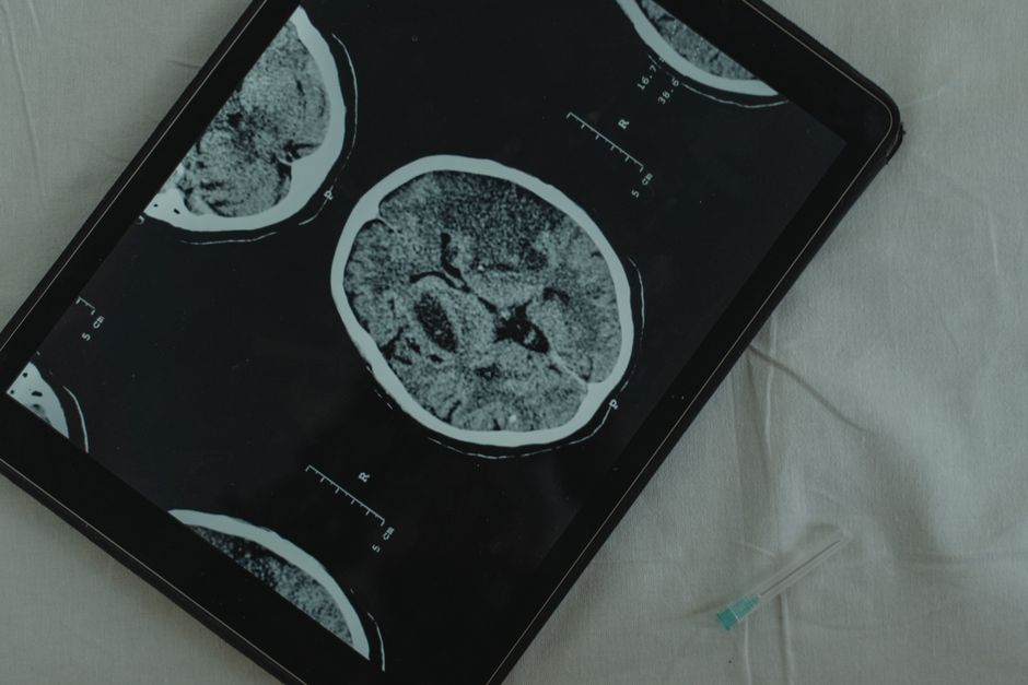
If your doctor sends you to a neurologist, the first question is often simple: what are they actually going to do to me? The answer starts with the neurological exam — a hands-on check of your reflexes, strength, sensation, coordination, and memory. From there, a neurologist chooses tests based on what that exam and your symptoms suggest. Those tests range from an EEG, which records the brain’s electrical activity, to imaging like CT, MRI, and PET scans, which show the brain’s structure and function. Not every patient needs every test. The goal is to answer a specific question about your nervous system.
What Is a Neurological Exam and Why Does It Come First?
The neurological exam is the foundation of everything that follows. It takes place in the office, requires no machines, and often tells the neurologist where to look before any scan is ordered.
A neurologist will typically assess several areas:
- Mental status — attention, memory, language, and orientation to time and place
- Cranial nerves — the twelve nerve pairs that control eye movement, facial strength, hearing, swallowing, and more
- Motor function — muscle strength and tone, tested against resistance
- Reflexes — including the knee and ankle jerks, plus the plantar reflex
- Sensation — light touch, pinprick, vibration, and position sense
- Coordination and gait — finger-to-nose testing, heel-to-shin testing, and how you walk
This exam is not a formality. It localizes the problem. A neurologist can often tell whether a symptom comes from the brain, the spinal cord, a nerve root, or a peripheral nerve before any imaging is done. That matters because it determines which test gets ordered next. Ordering an MRI of the brain when the problem is in the foot nerves wastes time and money.
What Does an EEG Measure and When Is It Used?
An EEG, or electroencephalogram, records the electrical activity of the brain. Small metal discs called electrodes are placed on the scalp, and the machine traces the brain’s electrical patterns onto a screen or paper.
The test is painless. It does not send any electricity into your head — it only listens. Most routine EEGs take about 20 to 40 minutes, though some are recorded over several hours or even days, especially when seizures are suspected but not captured in a short session.
EEGs are used most often for:
- Evaluating suspected seizures or epilepsy
- Looking at brain activity in someone with confusion or altered consciousness
- Monitoring brain function during certain surgeries
- Assessing sleep disorders in some cases
A normal EEG does not rule out epilepsy. Seizures can happen without showing up during a short recording, which is why longer monitoring is sometimes needed. This is one of the most common misunderstandings about the test.
EEG also does not measure intelligence, personality, or mental health. It records electrical patterns, nothing more.
What Do Brain Imaging Tests Like CT and MRI Show?
Imaging shows structure. CT and MRI are the two workhorses of neurological imaging, and they answer different questions.
A CT scan uses X-rays to create cross-sectional images of the brain. It is fast — often just a few minutes — and is the first choice in an emergency such as a suspected stroke or head injury, because it quickly shows bleeding, fractures, and large masses.
An MRI uses strong magnetic fields and radio waves, not radiation. It produces much more detailed images of brain tissue, the spinal cord, and nerves. MRI is better than CT for detecting small tumors, inflammation, multiple sclerosis lesions, and subtle changes in the brain’s white matter. A scan can take 30 to 60 minutes, and the machine is noisy and enclosed, which some people find difficult.
Both tests are generally safe for most people, but each has limits. CT involves radiation exposure, so it is used thoughtfully, especially in younger patients and pregnant women. MRI is not safe for people with certain metal implants, pacemakers, or metallic foreign bodies in the eye, because the magnetic field can move or heat them. You will be screened for these before an MRI.
Some MRI scans use a contrast dye containing gadolinium. This improves visibility of certain abnormalities. Gadolinium contrast is generally well tolerated, but it is not given to everyone, and kidney function may be checked first.





How Are CT and MRI Different From Each Other?
The two tests overlap but are not interchangeable. The table below shows the main differences.
| Feature | CT Scan | MRI |
|---|---|---|
| What it uses | X-rays | Magnetic fields and radio waves |
| Radiation | Yes | No |
| Typical time | A few minutes | 30 to 60 minutes |
| Best for | Emergency bleeding, fractures, fast answers | Detailed soft tissue, spinal cord, subtle lesions |
| Main limitation | Less detail in soft tissue | Not safe with certain metal implants |
Neither test is “better” in general. The right choice depends on the question being asked and the situation.
What Is a PET Scan and How Is It Different?
A PET scan, or positron emission tomography, shows how the brain is functioning rather than just how it looks. A small amount of a radioactive tracer is injected into the bloodstream. The tracer travels to the brain, and the scanner detects where it accumulates, revealing patterns of activity and metabolism.
PET is used in specific situations, not as a routine first test. It can help distinguish certain types of dementia, evaluate some brain tumors, and map brain activity in research settings. It is also used in some cases of epilepsy to locate the area where seizures begin when other tests are unclear.
PET scans involve radiation exposure and are more expensive and less widely available than CT or MRI. They are usually ordered when the answer cannot be found another way.
What Other Tests Do Neurologists Order?
Imaging and EEG are not the whole picture. Depending on the symptoms, a neurologist may also order:
- Nerve conduction studies and EMG — small electrical pulses and thin needle electrodes test how well nerves and muscles are working. These are used for conditions like carpal tunnel syndrome, neuropathy, and nerve injuries.
- Lumbar puncture (spinal tap) — a sample of cerebrospinal fluid is collected from the lower back and tested for infection, inflammation, or bleeding around the brain and spinal cord.
- Blood tests — these can reveal vitamin deficiencies, thyroid problems, infections, or autoimmune conditions that affect the nervous system.
- Evoked potentials — these measure how signals travel along nerve pathways in response to sight, sound, or touch.
- Sleep studies — used when sleep disorders or nighttime seizures are suspected.
Each test has a purpose. A neurologist does not order all of them at once. The sequence is guided by what the exam and your history suggest, and by what is most likely to answer the question at hand.
What Should You Expect During These Tests?
Most neurological tests are well tolerated. The neurological exam is a conversation and a series of simple movements. EEG electrodes are glued to the scalp with a paste that washes out afterward. CT scans are quick and quiet. MRI scans are loud but you are given earplugs or headphones, and you can usually communicate with the technologist during the scan.
Some tests are less comfortable. EMG needles can cause brief discomfort. A lumbar puncture involves a numbing shot and a period of lying flat afterward to reduce the risk of headache. PET scans require an injection and a waiting period before the scan itself.
If you are pregnant, tell your doctor before any test involving radiation, including CT and PET. MRI is generally avoided in the first trimester unless the benefit clearly outweighs the risk, though guidelines vary. If you have any metal implants, pacemakers, or a history of working with metal, tell the staff before an MRI.
Why Doesn’t Every Neurologist Order the Same Tests?
Two people with the same symptom can get different tests. That is not inconsistency — it reflects how neurology works. A headache with sudden onset and a headache that has been stable for years point in different directions. A seizure in a young adult and a seizure in someone over 65 raise different concerns.
The neurologist’s job is to narrow the possibilities efficiently. Sometimes that means starting with an EEG. Sometimes it means going straight to MRI. Sometimes it means watching and waiting, because the exam is normal and the symptoms are mild and stable.
Testing also has limits. A normal scan does not always mean nothing is wrong. Some conditions — certain headaches, some forms of epilepsy, and many functional neurological disorders — can produce real symptoms without showing structural changes on imaging. That does not make the symptoms less real. It means the test is not the right tool for that particular problem.
Frequently Asked Questions
What is the difference between an EEG and an MRI?
An EEG records the brain’s electrical activity over time, while an MRI creates detailed pictures of the brain’s structure. They answer different questions, and a neurologist may order one or both depending on your symptoms.
Does a normal EEG mean I don’t have epilepsy?
No. A normal EEG does not rule out epilepsy, because seizures may not occur during a short recording. Longer monitoring or other tests are sometimes needed to confirm or exclude the diagnosis.
Is an MRI or CT scan better for the brain?
Neither is better in general — they are used for different purposes. CT is faster and preferred in emergencies like suspected bleeding, while MRI gives more detail for soft tissue, the spinal cord, and subtle brain changes.
Are neurological tests painful?
Most are not. The neurological exam, EEG, CT, and MRI are generally painless. EMG needles and lumbar punctures can cause brief discomfort, and your care team can explain what to expect beforehand.
