
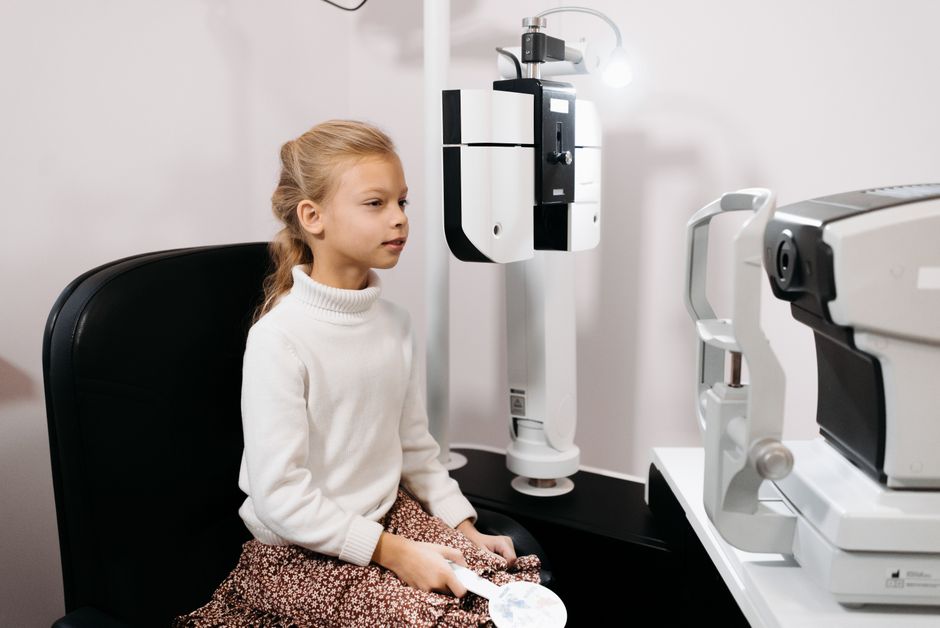
Ultrasound biomicroscopy (UBM) is a high-resolution imaging technique that uses high-frequency sound waves to create detailed pictures of the front part of the eye. It allows eye doctors to see structures that are hidden behind the iris or blocked by opaque tissue, which standard imaging cannot reach. UBM is most often used when a regular eye exam or other scans cannot provide enough information about the anterior segment — the cornea, iris, ciliary body, and lens.
What Is Ultrasound Biomicroscopy UBM In Ophthalmology?
UBM is an imaging method that produces cross-sectional images of the anterior segment of the eye using ultrasound frequencies typically in the range of 35 to 50 megahertz. That frequency is much higher than the 10 to 20 MHz used in conventional B-scan ocular ultrasound, which is why UBM produces finer detail but shallower penetration.
The trade-off is depth. UBM can image roughly the first 4 to 5 millimeters of tissue. That includes the cornea, anterior chamber, iris, ciliary body, and the anterior lens surface. Structures deeper in the eye — the vitreous, retina, and optic nerve — are better assessed with other tools.
Because sound waves travel through opaque tissue, UBM works even when there is a dense corneal scar, a hyphema (blood in the anterior chamber), or an opaque cornea. Optical methods like slit-lamp examination and anterior segment OCT cannot see through those obstacles. This is the core advantage of UBM.
How Does UBM Work Compared To Other Eye Imaging?
UBM uses a transducer that emits high-frequency sound waves and records the echoes that bounce back from tissue interfaces. A computer converts those echoes into a two-dimensional image. The probe is placed over the eye with a coupling medium — typically a saline-filled cup or gel — to maintain contact without pressing on the eye.
The resolution of UBM is approximately 20 to 50 micrometers, depending on the frequency used. That is fine enough to show individual ciliary processes and the zonules that hold the lens in place.
Compared to other imaging methods:
- Anterior segment OCT uses light waves and gives excellent resolution of the cornea and anterior chamber, but light cannot penetrate an opaque cornea or iris.
- Conventional B-scan ultrasound penetrates deeper but at lower resolution. It is better for the posterior segment.
- Slit-lamp examination is the standard first look, but it cannot see behind the iris or through dense scarring.
UBM fills a specific gap. It is not a replacement for any of these — it answers questions the others cannot.
What Conditions Can UBM Help Diagnose?
UBM is used in several clinical situations where the anterior segment anatomy needs detailed assessment. The evidence supporting its diagnostic value varies by condition.
Glaucoma assessment. UBM can show the configuration of the anterior chamber angle, the position of the ciliary body, and whether the iris is pushing forward against the trabecular meshwork. This is relevant in angle-closure glaucoma and in eyes where the angle cannot be seen clearly on gonioscopy. Some studies suggest UBM can identify plateau iris configuration, a variant where the ciliary body holds the peripheral iris forward even after a laser iridotomy.
Anterior segment tumors. UBM can help define the size, shape, and internal structure of iris and ciliary body tumors. It helps distinguish solid masses from cysts. However, UBM cannot determine whether a tumor is benign or malignant on its own — that requires clinical judgment and sometimes biopsy.
Trauma and foreign bodies. When the cornea is scarred or hazy after injury, UBM can locate retained foreign bodies and assess damage to the lens or ciliary body. This is one of its most established uses.
Implantable contact lens sizing. Before placing a phakic intraocular lens (an implantable lens for refractive correction), surgeons may use UBM to measure the distance between the lens and the posterior cornea. This helps reduce the risk of the implant touching the natural lens.
Ciliary body evaluation. In conditions like malignant glaucoma or post-surgical complications, UBM can show the position of the ciliary body and help guide treatment decisions.
The strength of evidence differs across these uses. For trauma and tumor localization, UBM is well established. For some glaucoma applications, it is widely used but the evidence base is more limited.





What Happens During A UBM Exam?
A UBM exam is performed in an eye clinic, usually by an ophthalmologist or a trained ophthalmic technician. The procedure takes about 10 to 20 minutes per eye.
The patient lies back or sits at a slit lamp. A topical anesthetic drop numbs the eye surface. A small plastic cup — called an eye cup or scleral shell — is placed between the eyelids and filled with a sterile coupling fluid, usually saline or methylcellulose. The ultrasound probe is then dipped into the fluid and moved across the eye without touching the surface directly.
This immersion technique is important. It keeps the probe at a safe distance and avoids putting pressure on the eye, which could distort the anatomy being measured.
During the scan, the patient may be asked to look in different directions so the doctor can capture views of the angle, ciliary body, and lens from multiple angles. The images appear on a screen in real time.
After the exam, the numbing drop wears off within 15 to 30 minutes. Most people can drive and return to normal activities right away. Some clinicians advise against rubbing the eye for a few hours.
Is UBM Safe? What Are The Risks?
UBM is generally considered a low-risk procedure. It uses sound waves, not radiation, so there is no ionizing radiation exposure. The ultrasound energy levels used in diagnostic UBM are within established safety limits for ocular imaging.
The main risks are related to the technique rather than the sound waves themselves:
- Corneal abrasion. The eye cup or probe can scratch the cornea if the eye moves suddenly. This is uncommon but possible. It usually heals within a day or two.
- Discomfort. Some people find the eye cup irritating. The topical anesthetic reduces this.
- Infection. Any device that contacts the eye carries a small risk of infection if not properly sterilized. Clinics follow standard disinfection protocols.
UBM should not be performed on an eye with an open globe injury unless specifically indicated and done with extreme care. In those cases, the risk of causing further damage must be weighed against the diagnostic benefit.
There are no known long-term effects from diagnostic ultrasound exposure to the eye at these frequencies and energy levels.
What Are The Limitations Of UBM?
UBM is not a general-purpose eye scan. It has clear boundaries.
It cannot image structures deeper than about 4 to 5 millimeters from the surface. That means the vitreous, retina, and optic nerve are outside its range. For those areas, doctors use B-scan ultrasound, OCT, or other methods.
UBM requires direct contact with the eye through a coupling medium. That makes it more operator-dependent than some other scans. The quality of the images depends heavily on the skill of the person performing the exam.
It also requires specialized equipment that not every eye clinic has. UBM machines are more common in academic centers, glaucoma specialty practices, and referral centers than in general optometry offices.
Finally, UBM produces cross-sectional images, not three-dimensional volumes like some newer imaging systems. The doctor must mentally reconstruct the anatomy from multiple two-dimensional slices.
How Does UBM Compare To Anterior Segment OCT?
Both UBM and anterior segment OCT image the front of the eye, but they work differently and have different strengths.
| Feature | UBM | Anterior Segment OCT |
|---|---|---|
| Energy source | High-frequency sound waves | Light waves (near-infrared) |
| Penetration through opaque tissue | Yes | No |
| Resolution | Approximately 20–50 micrometers | Approximately 3–10 micrometers |
| Imaging depth | About 4–5 mm | Limited by light penetration; typically less through scarring |
| Contact required | Yes (immersion technique) | No (non-contact) |
| Best for | Behind iris, opaque cornea, ciliary body | Cornea, anterior chamber, clear media |
In plain terms: OCT gives sharper pictures when the eye is clear. UBM gives pictures when the eye is not clear, or when the area of interest is behind the iris. Many clinics use both depending on the question being asked.
When Would A Doctor Recommend UBM?
A doctor might recommend UBM when a standard exam leaves an important question unanswered. Common reasons include:
- Suspected angle-closure glaucoma where the angle cannot be seen clearly
- A mass behind the iris that needs to be measured or characterized
- History of eye trauma with retained foreign body or hazy cornea
- Planning for certain types of eye surgery, such as phakic lens implantation
- Unexplained shallowing of the anterior chamber after surgery
UBM is not a routine screening test. It is ordered when the clinical picture calls for a detailed look at structures that other tools cannot reach. If your eye doctor recommends it, the reason is usually specific to your situation.
Some clinicians use UBM in research settings to study accommodation, the process by which the eye changes focus. That is an active area of investigation, not yet part of standard clinical care.
Frequently Asked Questions
Does UBM hurt?
UBM is generally not painful. The eye is numbed with drops, and the probe does not touch the eye directly — it sits in a fluid-filled cup. Some people feel mild pressure or irritation from the eye cup itself.
How long does a UBM exam take?
The scan usually takes about 10 to 20 minutes per eye. Including preparation and after-care, you can expect to be in the clinic for 30 to 45 minutes.
Can UBM see the retina?
No. UBM images only the front 4 to 5 millimeters of the eye. The retina and vitreous are too deep to be seen with this technique. A different type of ultrasound or OCT is used for those structures.
Is UBM better than OCT for glaucoma?
Neither is universally better. UBM can see through opaque tissue and image the ciliary body, which OCT cannot. OCT gives higher-resolution images when the cornea and anterior chamber are clear. The choice depends on what the doctor needs to see.
