
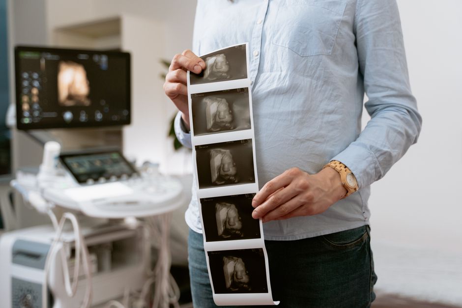
Ultrasound imaging uses high-frequency sound waves to create pictures of structures inside the body. A device called a transducer sends sound waves in and listens for the echoes that bounce back. A computer turns those echoes into real-time images. There is no ionizing radiation involved, which is one reason it is used so widely in pregnancy, heart care, and emergency medicine. It is also one of the most operator-dependent imaging tools in medicine — the skill of the person holding the probe matters a great deal.
What Is Ultrasound Imaging and How Does It Work?
Ultrasound — also called sonography — works on the same basic principle as a bat navigating in the dark. The transducer emits sound waves at frequencies far above what human ears can detect, typically in the range of 2 to 18 megahertz. These waves travel through tissue and bounce back at different speeds depending on the density of what they hit. Fluid reflects very little. Bone reflects almost everything.
The transducer alternates between sending pulses and receiving echoes, thousands of times per second. A processor measures how long each echo takes to return and how strong it is, then maps those signals into a two-dimensional image. Because this happens continuously, the image moves in real time. That is a key difference from X-ray or CT, which capture a single moment.
Different tissues produce different echo patterns:
- Fluid (blood, urine, amniotic fluid) appears black — it is anechoic, meaning it reflects almost no sound.
- Soft tissue (liver, kidney, muscle) appears in shades of gray.
- Bone and dense structures appear bright white and cast shadows behind them.
- Air scatters sound waves, which is why ultrasound does not image the lungs well.
That last point explains a lot about where ultrasound works and where it does not. Sound needs a medium to travel through. Air and bone disrupt the signal. This is why ultrasound excels at imaging soft tissue, organs, and fluid-filled spaces but struggles with lungs and areas behind bone.
What Are the Main Uses of Ultrasound Imaging?
Ultrasound is used across nearly every medical specialty. Some uses are diagnostic. Others guide procedures in real time. A few of the most established applications include:
Obstetrics and pregnancy. Ultrasound is the standard tool for monitoring fetal development, estimating gestational age, checking placenta position, and detecting certain abnormalities. It is also used to guide amniocentesis and other prenatal procedures.
Cardiology. An echocardiogram shows the heart’s chambers, valves, and wall motion. It can measure ejection fraction — the percentage of blood pumped out of the left ventricle with each beat. A normal ejection fraction is generally between 50% and 70%, though this range can vary slightly depending on the reference lab and the method used.
Abdominal imaging. Ultrasound is often the first test for gallbladder disease, kidney stones, liver conditions, and appendicitis. It is fast, portable, and does not require contrast dye.
Vascular imaging. Doppler ultrasound measures blood flow through arteries and veins. It is commonly used to check for deep vein thrombosis and carotid artery narrowing.
Musculoskeletal and soft tissue. Tendons, ligaments, muscles, and joints can be examined for tears, inflammation, or fluid buildup. Ultrasound is also used to guide injections into specific joints or tissues.
Emergency and critical care. Focused ultrasound exams help clinicians quickly assess for internal bleeding, collapsed lungs, or cardiac activity during resuscitation. This use has expanded significantly over the past two decades.
Thyroid and breast imaging. Ultrasound helps characterize nodules found on physical exam or other imaging. It is often used to guide biopsies of suspicious tissue.
What Are the Different Types of Ultrasound?
Not all ultrasound exams work the same way. The type used depends on what the clinician needs to see.
2D grayscale ultrasound is the standard. It produces flat, cross-sectional images and is what most people picture when they think of an ultrasound.
Doppler ultrasound detects movement — usually blood flow. It can show direction and speed. Color Doppler overlays color onto the image to show flow direction. Power Doppler is more sensitive to slow flow but does not show direction.
3D and 4D ultrasound build a three-dimensional volume from many 2D slices. 4D adds motion, showing the volume in real time. These are most familiar in pregnancy imaging. While they can provide detailed views of fetal anatomy, they are not routinely required for standard prenatal care. Some clinicians use them for specific diagnostic questions.
Elastography measures tissue stiffness. It is used primarily in liver imaging to assess fibrosis and in breast imaging to help characterize lesions. Stiffer tissue often indicates disease, though the relationship is not absolute.
Contrast-enhanced ultrasound uses microbubble contrast agents to improve visualization of blood flow in organs like the liver. This technique is more established in Europe and Canada than in the United States, where its use is growing but still limited.
Point-of-care ultrasound refers to ultrasound performed at the bedside by the treating clinician — often in emergency rooms, intensive care units, and outpatient clinics. It answers focused questions quickly rather than providing a full diagnostic study.




Is Ultrasound Safe?
Ultrasound does not use ionizing radiation, unlike X-rays and CT scans. That is a genuine advantage. It also has no known cumulative dose limit for diagnostic use, which is why it can be repeated as needed.
That said, “no ionizing radiation” does not mean “no biological effect whatsoever.” Ultrasound waves carry energy. At high intensities, that energy can heat tissue slightly and, in laboratory settings, produce mechanical effects in cells. Diagnostic ultrasound operates at intensities well below the levels where these effects have been observed in humans. Decades of use have not produced evidence of harm at standard diagnostic settings.
The safety picture is well established for diagnostic ultrasound used by trained professionals for medical indications. It is less clear for prolonged, non-medical use — such as keepsake fetal imaging performed without a medical reason, sometimes for extended sessions. Professional bodies generally advise against this practice because the risk-benefit calculation changes when there is no diagnostic benefit.
For standard diagnostic exams, the benefits almost always outweigh the theoretical risks. The key qualifier is “standard diagnostic” — meaning performed by a trained operator, for a medical reason, at settings consistent with professional guidelines.
What Should You Expect During an Ultrasound Exam?
Most ultrasound exams are straightforward and take between 15 and 45 minutes, depending on the area being studied.
For many abdominal and pelvic exams, you may be asked to arrive with a full bladder. A full bladder pushes other structures out of the way and gives the sound waves a clear path. For other exams, you may be asked to fast beforehand. Instructions vary by the specific study, so follow whatever your provider gives you.
You will lie on an exam table. The technologist applies a clear gel to your skin. The gel eliminates air between the transducer and your skin, which would otherwise block the sound waves. The transducer is pressed against your skin and moved around. You may feel mild pressure but should not feel pain. If you do, say so.
For certain exams — such as transvaginal or transesophageal ultrasound — the transducer is placed inside the body. These are performed when a clearer view is needed than what external scanning can provide. They are generally well tolerated, though they may be uncomfortable for some people.
After the exam, a radiologist or other qualified clinician reviews the images and sends a report to your referring provider. Results are typically available within a few days, though urgent findings are communicated sooner.
What Are the Limitations of Ultrasound?
Ultrasound has real limitations that are worth understanding.
It cannot see through bone or air. That means it cannot image the brain through an intact skull in adults, and it cannot provide detailed images of the lungs. It also has limited depth penetration — deeper structures are harder to resolve clearly.
Image quality depends heavily on the operator. Two technologists scanning the same patient can produce different images. This is not a flaw unique to ultrasound, but it is more pronounced than with CT or MRI, where the machine does more of the work.
Body habitus matters. More tissue between the transducer and the target structure means more signal attenuation. This can make images harder to interpret in patients with higher body mass index, though advances in transducer technology have improved this to some degree.
Ultrasound is also not a replacement for other imaging when those are indicated. A normal ultrasound does not rule out every condition. If a clinician suspects a problem that ultrasound cannot adequately assess, they may order a CT, MRI, or other study.
How Does Ultrasound Compare to Other Imaging Methods?
Each imaging method has trade-offs. The table below summarizes the main differences.
| Method | Ionizing Radiation | Best For | Main Limitation |
|---|---|---|---|
| Ultrasound | No | Soft tissue, fluid, real-time movement, pregnancy | Cannot penetrate bone or air; operator-dependent |
| X-ray | Yes | Bone, chest, quick screening | Limited soft tissue detail; radiation exposure |
| CT | Yes | Detailed cross-sections, trauma, cancer staging | Higher radiation dose than X-ray |
| MRI | No | Brain, spine, joints, soft tissue detail | Expensive, slower, not for patients with certain implants |
The choice among these is a clinical decision. Ultrasound is often first because it is fast, portable, inexpensive, and safe. When it cannot answer the question, other methods step in.
Frequently Asked Questions
Is ultrasound imaging safe during pregnancy?
Diagnostic ultrasound has been used in pregnancy for decades without evidence of harm at standard settings. Professional guidelines recommend it be performed only when medically indicated, rather than for keepsake purposes.
Does ultrasound use radiation?
No. Ultrasound uses high-frequency sound waves, not ionizing radiation. This is a key reason it is preferred for pregnancy and for repeated exams.
How long does an ultrasound exam take?
Most exams take between 15 and 45 minutes, depending on the area being studied. Some focused point-of-care exams take only a few minutes.
Can ultrasound detect cancer?
Ultrasound can identify suspicious masses and guide biopsies, but it cannot definitively diagnose cancer on its own. A tissue sample examined under a microscope is generally required for a cancer diagnosis.
