
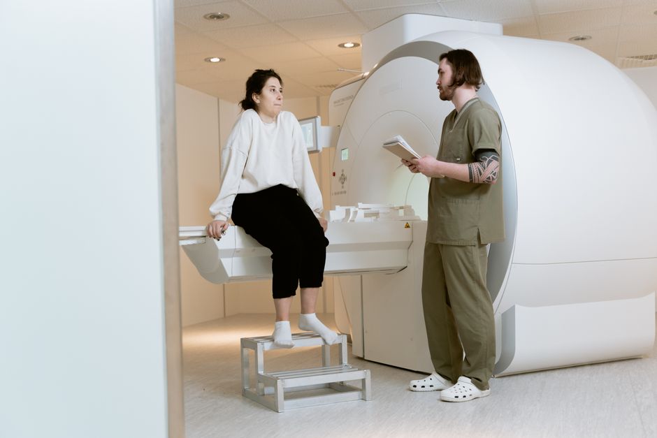
A CT scan is a medical imaging test that uses X-rays and a computer to create detailed cross-sectional pictures of the inside of your body. CT enterography is a specialized version of this scan that focuses specifically on the small intestine. It involves drinking a contrast liquid and receiving an IV contrast dye to make the bowel walls and any abnormalities stand out clearly on the images. This combination allows doctors to see inflammation, bleeding, tumors, and other problems in the small bowel that standard CT scans often miss.
What Is a CT Scan and How Does CT Enterography Work?
A CT scan, which stands for computed tomography, takes many X-ray images from different angles around your body. A computer then combines these images to create slices or cross-sections. Think of it like looking at a loaf of bread one slice at a time rather than just looking at the whole loaf from the outside. This gives doctors a much clearer view of your organs, bones, and blood vessels.
CT enterography builds on this basic technology but tailors it for the small intestine. The small bowel is long and coiled, making it hard to see with regular imaging. Before the scan, you drink about one to two liters of a neutral contrast liquid over 45 to 60 minutes. This liquid distends or stretches the bowel walls so they open up like a deflated balloon being filled with air. Without this distention, the walls collapse against each other and hide any problems.
You also receive an IV injection of iodinated contrast dye right before the scan. This dye travels through your bloodstream and highlights areas of inflammation or abnormal blood flow. The entire scanning process itself takes less than 30 seconds while you hold your breath. The images are then reconstructed to show the bowel wall in fine detail. Research published in the American Journal of Roentgenology has shown that CT enterography detects active inflammation in Crohn’s disease with over 90% accuracy.
Who Needs This Scan and What Conditions Does It Detect?
Doctors most commonly order CT enterography for people with suspected or known Crohn’s disease. This chronic inflammatory bowel disease causes swelling, ulcers, and narrowing in the small intestine. A standard colonoscopy can only reach the lower part of the small bowel, leaving the upper two-thirds invisible to the scope. CT enterography fills that gap by imaging the entire small intestine.
Other conditions this scan can detect include small bowel tumors, both cancerous and benign. It can identify sources of gastrointestinal bleeding that other tests have missed. It also detects obstructions where the bowel is blocked, fistulas which are abnormal connections between the bowel and other organs, and abscesses which are pockets of infection. The scan can show complications from radiation therapy or surgery in the abdomen.
Some patients with unexplained abdominal pain, weight loss, or chronic diarrhea may also be candidates if standard tests have been inconclusive. The American College of Radiology recommends CT enterography as a first-line imaging test for suspected small bowel Crohn’s disease. It is not typically used for routine screening. It is a problem-solving tool for when symptoms point to the small intestine but other tests have not found the cause.
How Do You Prepare for a CT Enterography Scan?
Preparation is straightforward but requires some planning. You need to stop eating solid food for about four hours before the scan. You can usually drink clear liquids like water, black coffee, or apple juice during this time. The contrast liquid you drink before the scan counts as a clear liquid.
You must tell your doctor about any medications you take, especially metformin for diabetes. Metformin can interact with the IV contrast dye and cause a rare but serious kidney problem. Your doctor may ask you to stop taking it for 48 hours after the scan. You also need to inform the staff if you have any allergies, particularly to iodine or shellfish, since the contrast dye contains iodine.
Kidney function is checked before the scan because the contrast dye is filtered through your kidneys. If your kidney function is poor, the dye can cause further damage. In those cases, the doctor may choose a different imaging method. You should also tell the technologist if there is any chance you could be pregnant. CT scans use radiation, and while the dose is low, it is avoided during pregnancy unless absolutely necessary.
Wear comfortable clothing without metal zippers or buttons. You will likely change into a hospital gown anyway. Remove jewelry, glasses, and dentures before the scan. The entire appointment takes about 90 minutes total, with most of that time spent drinking the contrast liquid and waiting for it to move through your bowel.


What Are the Risks and Side Effects of CT Enterography?
The main risk from any CT scan is exposure to ionizing radiation. CT enterography delivers a radiation dose roughly equivalent to what you would receive from natural background radiation over one to three years. That is about 5 to 10 millisieverts. For comparison, a chest X-ray is about 0.1 millisieverts. The risk from a single scan is very small, but the lifetime risk increases with multiple scans. The American College of Radiology advises that the benefit of an appropriately ordered scan almost always outweighs this small risk.
The IV contrast dye carries its own risks. Some people experience a warm sensation or a metallic taste in the mouth as the dye is injected. These sensations pass within seconds. Allergic reactions to the dye occur in about 1 in 1000 people. Most reactions are mild, like hives or nausea. Severe reactions that cause trouble breathing are rare, happening in about 1 in 10,000 to 1 in 50,000 people. The radiology department is equipped to handle these emergencies.
Contrast-induced kidney injury is another concern. This happens when the dye damages kidney tissue, usually in people who already have reduced kidney function. The risk is about 2% in people with normal kidneys and higher in those with diabetes or pre-existing kidney disease. This is why your creatinine level is checked before the scan. Drinking plenty of water after the scan helps flush the dye out of your system.
The oral contrast liquid can cause bloating, cramping, or diarrhea. Some people find it difficult to drink the full amount because it is a large volume. Nausea is also possible. These side effects are temporary and resolve once the liquid passes through your system.
How Does CT Enterography Compare to Other Small Bowel Imaging Tests?
There are several ways to image the small intestine, and each has strengths and weaknesses. The table below compares CT enterography with the most common alternatives.
| Test | What It Shows Best | Limitations |
|---|---|---|
| CT Enterography | Bowel wall thickness, inflammation, fistulas, abscesses | Radiation exposure, need for IV contrast |
| MR Enterography | Same as CT but with better soft tissue detail | No radiation, longer scan time, more expensive |
| Capsule Endoscopy | Direct view of the lining of the small bowel | Cannot see through wall, risk of capsule getting stuck |
| Small Bowel Follow-Through | Barium outlines the bowel lumen | Lower detail, longer test, more radiation than CT |
MR enterography is very similar to CT enterography but uses magnetic fields instead of radiation. It is preferred for younger patients and those who need repeated scans because there is no radiation exposure. The trade-off is that MR scans take longer, often 30 to 45 minutes, and require you to hold still. Some people find the enclosed MRI machine uncomfortable.
Capsule endoscopy involves swallowing a tiny camera that takes pictures as it travels through your digestive tract. It provides a direct view of the bowel lining but cannot see through the wall. It also carries a small risk of the capsule becoming stuck at a narrowed area, which then requires surgical removal. CT enterography is usually done first because it can detect these narrowings before a capsule is used.
What Happens After the Scan and How Do You Get Results?
After the scan is complete, you can return to your normal activities immediately. There are no restrictions. You should drink extra water for the rest of the day to help your kidneys clear the contrast dye. If you are taking metformin, your doctor will tell you when it is safe to restart it, usually after 48 hours.
A radiologist, who is a doctor specialized in interpreting medical images, will review your scan. They look for areas of bowel wall thickening, abnormal enhancement from the contrast dye, narrowed segments, fistulas, abscesses, and masses. They also check the other organs visible in the scan, such as the liver, pancreas, kidneys, and spleen, for any incidental findings.
The results are typically available within 24 to 48 hours. Your ordering doctor will discuss the findings with you and explain what they mean for your condition. If the scan shows active inflammation, your doctor may adjust your medications or recommend further treatment. If it shows a stricture or narrowing, you may need a procedure to open it. If it shows a tumor, you will likely need a biopsy to determine if it is cancerous.
One thing many people do not realize is that a normal CT enterography does not completely rule out all small bowel disease. Very small ulcers or early inflammation may not be visible. If your symptoms continue despite a normal scan, your doctor may recommend capsule endoscopy or another test to look more closely at the bowel lining.
Frequently Asked Questions
Frequently Asked Questions
How long does a CT enterography scan take?
The actual scan takes less than 30 seconds, but the entire appointment lasts about 90 minutes because you need to drink the contrast liquid beforehand.
Is CT enterography painful?
No, the scan itself is painless. You may feel a warm sensation from the IV contrast injection and some bloating from the oral contrast liquid.
Can I eat before a CT enterography scan?
You should stop eating solid food four hours before the scan, but you can usually drink clear liquids including the contrast liquid you are given.
Does insurance cover CT enterography?
Most insurance plans cover CT enterography when it is ordered by a doctor for a medically necessary reason such as suspected Crohn’s disease or unexplained bleeding.
