
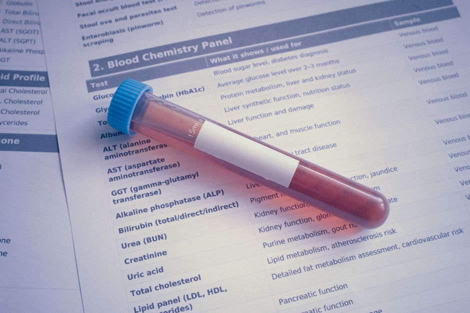
Cell viability is a measure of how many living cells are present in a sample at a given time. It is expressed as a percentage of the total cell population that is alive and functional. Scientists measure it using methods that detect biological markers unique to living cells, such as metabolic activity, membrane integrity, or ATP production.
What Does Cell Viability Mean And How Is It Measured?
Cell viability simply means the number of live cells in a population. A sample with high viability has mostly healthy, living cells. A sample with low viability has many dead or dying cells.
Researchers measure viability because it tells them whether a treatment works, whether cells are healthy, or whether a product is safe. The methods used to measure it fall into two broad categories: direct counting and indirect detection.
Direct counting uses a microscope to count live and dead cells. Indirect methods use chemical dyes or biological markers that react differently with living and dead cells. Both approaches are standard in research laboratories.
Why Does Cell Viability Matter in Research?
Cell viability is a core measurement in drug development, toxicology, and cancer research. When a new drug is tested, researchers need to know if it kills cancer cells without harming healthy cells. Viability assays provide that answer.
Viability also matters for quality control. Cell cultures used in manufacturing, such as vaccine production or regenerative medicine, must be healthy before use. A low viability reading can halt an entire production run.
In clinical diagnostics, viability testing helps monitor conditions like fertility. Sperm viability testing, for example, is a standard part of fertility evaluations. The same principles apply across all these fields.
What Are the Main Methods for Measuring Cell Viability?
Several methods exist, and each works on a different biological principle. The choice of method depends on the cell type, the equipment available, and the question being asked.
- Trypan blue exclusion: A dye enters dead cells but cannot pass through the intact membrane of living cells. Live cells appear clear; dead cells appear blue under a microscope.
- MTT assay: Living cells convert a yellow compound into a purple crystal. The amount of color produced correlates with the number of metabolically active cells.
- ATP measurement: Living cells contain ATP, the energy molecule. Dead cells quickly lose ATP. Measuring ATP levels gives a direct readout of viable cell number.
- Fluorescent dyes: Certain dyes bind to DNA only in dead cells. Others are converted by live-cell enzymes into a fluorescent signal. These can be read by specialized plate readers.
Each method has strengths and limitations. Trypan blue is simple and cheap but requires a skilled eye. The MTT assay is reliable but requires cell lysis, so the same cells cannot be used again. ATP assays are highly sensitive but cost more per sample.



How Do Viability and Proliferation Differ?
Viability and proliferation are often confused, but they measure different things. Viability tells you whether cells are alive right now. Proliferation tells you how fast cells are dividing and increasing in number.
Cells can be alive but not dividing. This happens in resting states, such as mature neurons or certain immune cells. A viability assay would show these cells as alive. A proliferation assay would show no change in number.
Some assays measure both simultaneously. For example, a live-cell imaging system can track cell number over time. This gives a fuller picture of what a treatment actually does to a cell population.
What Are the Limitations of Viability Assays?
No single viability assay is perfect. Each one can produce misleading results under certain conditions.
Metabolic assays like MTT depend on cellular enzymes. If a treatment changes the activity of those enzymes without killing cells, the assay can report false low viability. The cells are alive, but the assay reads them as dead.
Membrane integrity assays like trypan blue can miss early stages of cell death. Cells may have damaged internal machinery but still have an intact membrane. These cells stain as alive even though they are not functional.
Researchers often run two different types of assays to confirm results. If both methods agree, the conclusion is stronger. If they disagree, additional investigation is needed.
What Is the Difference Between Cell Viability and Cell Toxicity?
These terms are related but not identical. Viability measures how many cells survive. Toxicity measures how harmful a substance is to cells.
A highly toxic substance will reduce viability because it kills cells. But a substance can reduce viability without being directly toxic. For example, a drug might slow metabolism so much that cells cannot maintain basic functions, leading to death over time.
Toxicity assays usually expose cells to a substance at different concentrations and then measure viability. The result is often expressed as an IC50 value, which is the concentration that reduces viability by half. This number allows researchers to compare the potency of different substances.
How Is Cell Viability Measured in Clinical Settings?
In clinical laboratories, viability testing is used for specific diagnostic purposes. Sperm viability testing is common in fertility clinics. White blood cell viability matters in transfusion medicine. Tumor cell viability is assessed in some cancer treatment monitoring protocols.
Flow cytometry is the gold standard in many clinical settings. This instrument passes cells through a laser beam one at a time. Fluorescent markers on the cells emit light that the machine detects. It can count thousands of cells in seconds and distinguish live from dead with high precision.
Flow cytometry is more expensive and requires specialized training. But it provides data that simple counting cannot match, including information about cell size, shape, and surface markers.
Can Cell Viability Assays Be Used at Home?
No commercial home test currently measures cell viability for consumer health purposes. The equipment and reagents required are designed for laboratory use.
Some wellness companies market tests that claim to measure cellular health or vitality. These products are not the same as cell viability assays. No clinical evidence confirms that consumer tests can meaningfully measure cell viability in a way that guides health decisions.
If you are considering a product that claims to measure your cells’ health, treat the claim with caution. Ask what specific biomarker the test measures and whether that biomarker has been validated in peer-reviewed studies.
Frequently Asked Questions
What is the simplest way to measure cell viability?
Trypan blue exclusion with a hemocytometer is the simplest and most accessible method. It requires only a microscope, a counting chamber, and the dye itself.
How long does a cell viability assay take?
Simple methods like trypan blue take about 10 to 15 minutes from sample to result. Metabolic assays like MTT typically require 2 to 4 hours because they depend on enzyme reactions and color development.
Can cell viability be measured in living tissue?
Yes, some techniques can assess viability in intact tissue, but they are more complex. Fluorescent dyes that penetrate tissue and specialized imaging systems are used in research settings, not routine clinical practice.
What does a viability percentage of 90% mean?
It means 90 out of every 100 cells in the sample are alive. In most cell culture work, a viability of 90% or higher is considered healthy and acceptable for experimental use.
