
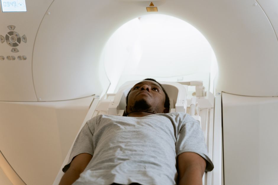
A CT scan is a three-dimensional X-ray study that shows the inside of the body in cross-sections, slice by slice. Doctors use it to diagnose problems in nearly every part of the body — broken bones, bleeding in the brain, tumors, kidney stones, blood clots in the lungs, and infections deep inside the abdomen. It is one of the most versatile imaging tools in medicine, and it can answer questions that plain X-rays and ultrasounds often cannot.
What Do CT Scans Diagnose?
CT scans diagnose conditions that involve structure — something that is broken, blocked, bleeding, swollen, torn, or growing where it should not be. The scan does not diagnose everything, though. It shows anatomy extremely well and function less well.
Common categories include:
- Trauma: Bleeding inside the skull, a torn spleen, a collapsed lung, or a fractured vertebra after an accident.
- Cancers and tumors: The size, location, and spread of tumors in the chest, abdomen, pelvis, and elsewhere.
- Blockages: Kidney stones, gallstones, bowel obstruction, or a blocked artery.
- Infections: Abscesses, infected fluid collections, pneumonia, and sinus infections.
- Blood vessel problems: Aneurysms, blood clots in the lungs, and tears in the wall of the aorta.
CT is often the fastest way to get a clear answer in an emergency. A person with sudden severe abdominal pain, a head injury, or signs of a stroke may get a CT within minutes of arriving at a hospital.
How Does a CT Scan Actually Work?
A CT scanner is a large ring-shaped machine. The patient lies on a table that slides through the center of the ring. Inside, an X-ray tube and detectors spin rapidly around the body, taking hundreds of images from many angles.
A computer then combines those images into cross-sectional pictures. Instead of one flat shadow like a standard X-ray, you get a stack of thin slices that can be viewed from the front, side, top, or any angle. That is why CT is sometimes called “CAT scan” — computed axial tomography.
Some scans use a contrast dye, given by mouth, injected into a vein, or both. Contrast makes certain tissues and blood vessels show up more clearly. Not every scan needs it, and the decision depends on what the doctor is looking for.
What Can a CT Scan Show That Other Tests Cannot?
CT’s main advantage is speed and detail at the same time. An ultrasound is excellent for the gallbladder, kidneys, and unborn babies but cannot see through bone or air. An MRI gives superb soft-tissue detail but takes longer and is harder to use in an unstable patient.
CT sits in a useful middle ground. It images bone, air, soft tissue, and blood vessels in one pass, and it does so in seconds to minutes. For a patient who is bleeding internally, that speed matters.
Another advantage is that CT images can be reconstructed in three dimensions. A surgeon planning to remove a tumor can see exactly how it sits relative to nearby organs and blood vessels before making a single cut.





Where in the Body Are CT Scans Used?
CT is used from head to toe. The table below lists some common areas and what a scan of that area typically looks for.
| Body Area | Common Reasons for a CT Scan |
|---|---|
| Head | Bleeding after injury, stroke, sinus infection, skull fracture |
| Chest | Blood clot in the lungs, lung cancer, pneumonia, aortic problems |
| Abdomen and pelvis | Appendicitis, kidney stones, tumors, abscess, bowel blockage |
| Spine | Fractures, disc problems, narrowing of the spinal canal |
| Heart | Calcium buildup in coronary arteries, sometimes blockages |
| Bones and joints | Complex fractures, bone tumors, joint damage |
One clarification worth making: a CT scan of the chest does not “screen for everything.” It is a targeted test. What it can see depends on whether contrast was used and how the scan was set up.
What Does a CT Scan Not Diagnose Well?
CT is not the best tool for every question. Being honest about its limits helps you understand why a doctor might order a different test.
CT does not image the spinal cord, nerves, or brain tissue as clearly as MRI. It cannot reliably show early changes in the brain from a stroke in the first hours — it mainly rules out bleeding. It is also not the first choice for the gallbladder or the ovaries in many cases, where ultrasound tends to be preferred.
CT also cannot tell you what a tissue is made of at the cellular level. A mass that looks suspicious on CT usually needs a biopsy to confirm whether it is cancer, and what type.
Is the Radiation From a CT Scan a Concern?
CT scans use ionizing radiation, and the dose is higher than a standard X-ray. That is a real consideration, not a marketing point.
The benefit usually outweighs the risk when a scan is medically needed — for example, to find internal bleeding or diagnose a life-threatening clot. But the risk is not zero, and it accumulates with repeated scans over a lifetime.
This is why doctors generally do not order CT scans casually. When a condition can be diagnosed another way — with ultrasound, MRI, or a physical exam — those options are often considered first. If you are concerned about radiation, it is reasonable to ask your doctor whether a different test could answer the same question.
What Happens During and After a CT Scan?
Most CT scans take only a few minutes. You may be asked to lie still, sometimes hold your breath briefly. If contrast is used, you may feel a warm flush or a metallic taste — these sensations pass quickly.
Results are read by a radiologist, who sends a report to your doctor. Some findings are urgent and reported immediately. Others are routine and discussed at your next appointment.
Occasionally a scan shows an unexpected finding — something unrelated to the original question, sometimes called an incidental finding. Many of these turn out to be harmless, but some require follow-up testing. Your doctor will explain what, if anything, needs to happen next.
How Does CT Compare to MRI and Ultrasound?
Each imaging test has strengths. The right choice depends on the body part, the question being asked, and the patient’s situation.
- CT: Fast, excellent for bone and acute bleeding, uses radiation, often needs contrast.
- MRI: Best for soft tissue, brain, spinal cord, and joints, no radiation, but slower and not ideal for unstable patients.
- Ultrasound: No radiation, real-time imaging, good for gallbladder, kidneys, and pregnancy, but limited by bone and air.
In an emergency, speed often decides the choice. In a non-urgent situation, a doctor may pick the test that gives the clearest answer with the least risk.
Frequently Asked Questions
What conditions can a CT scan detect?
A CT scan can detect fractures, internal bleeding, tumors, blood clots, kidney stones, infections, and blockages in many parts of the body. It is especially useful in emergencies because it provides detailed images quickly.
Can a CT scan detect cancer?
CT scans can show masses or abnormal tissue that may be cancer, and they help determine a tumor’s size and location. However, a biopsy is usually needed to confirm whether a mass is actually cancer.
Does a CT scan show everything in the body?
No, a CT scan shows structure well but has limits. It does not image nerves, the spinal cord, or brain tissue as clearly as MRI, and it cannot show what a tissue is made of at the cellular level.
Is a CT scan safe?
CT scans use ionizing radiation, which carries some risk, especially with repeated scans over time. When a scan is medically necessary, doctors generally consider the benefit to outweigh the risk.
