
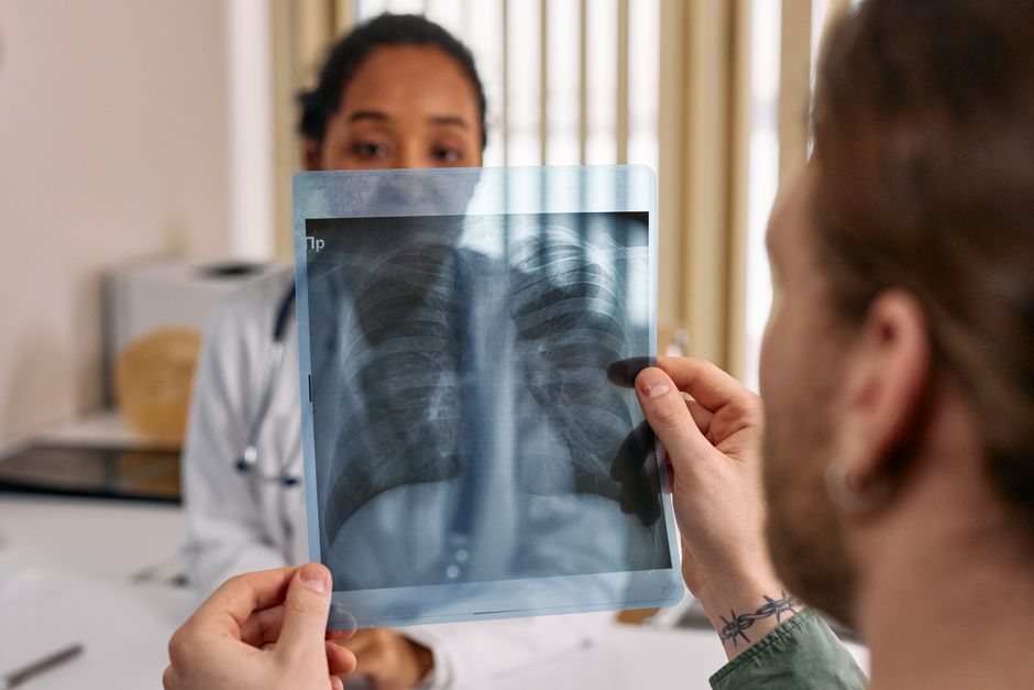
A chest X-ray is a two-dimensional shadow image of everything between your lungs and your breastbone. Dense structures like bone and fluid block more X-ray beams and show up white. Air-filled spaces like healthy lungs let beams pass through and show up black. That single fact explains most of what a chest X-ray can and cannot tell a doctor.
It is one of the most common imaging tests in medicine. It is fast, low-cost, and exposes you to a small amount of radiation. In many situations it is the first picture a clinician reaches for when something is wrong in the chest.
What Do Chest X Rays Reveal?
A chest X-ray can reveal pneumonia, a collapsed lung, fluid around the lungs, an enlarged heart, a mass or nodule, and certain bone problems in the chest wall. It can also show free air under the diaphragm, which is a sign of a perforated bowel.
What it often cannot do is give a precise diagnosis on its own. A shadow on an X-ray is a finding, not a disease. The same white patch can come from pneumonia, a tumor, bleeding, or scar tissue. Doctors combine the image with your symptoms, your history, and sometimes follow-up scans to figure out what it means.
This is the core limitation to understand. A chest X-ray is a screening and triage tool. It points toward an answer. It frequently does not deliver the final one.
What Can a Chest X-Ray Show in the Lungs?
The lungs are the main reason most chest X-rays get ordered. Because air is nearly transparent to X-rays, healthy lung tissue appears dark. Anything that replaces air with fluid, cells, or scar tissue appears whiter.
That principle lets a radiologist spot several patterns:
- Consolidation — areas where air sacs fill with fluid or cells, often from pneumonia
- Effusion — fluid collecting in the space between the lung and chest wall
- Pneumothorax — air trapped around the lung, which can cause it to collapse
- Nodules or masses — round or irregular densities that may be benign or may need further testing
- Scarring — old areas of damage that look similar to active disease
Pneumonia is one of the most common findings. But here is a point many people miss: a chest X-ray can look normal early in an infection, especially in people who are dehydrated or whose immune system is not mounting a strong response. A normal film does not always rule out pneumonia. Some clinicians repeat the image a day or two later if symptoms persist.
Small nodules are another area where the X-ray has limits. A nodule can be too small to see on a standard chest film, or it can be hidden behind a rib, the heart, or the diaphragm. When doctors need a closer look at lung tissue, they usually turn to a CT scan, which takes many cross-sectional images instead of one flat shadow.
What Does a Chest X-Ray Show About the Heart and Blood Vessels?
A chest X-ray can show whether the heart looks enlarged. The heart sits in the middle of the chest, and its silhouette is measured against the width of the rib cage. If the heart shadow takes up more than about half the chest width on a standard frontal view, doctors call it an enlarged cardiac silhouette.
An enlarged silhouette is a clue, not a diagnosis. It can mean a genuinely enlarged heart. It can also mean fluid around the heart, or simply that the person was lying down or breathed in poorly when the image was taken. A poor-quality film can mimic disease, which is why the technical quality of the image matters so much.
The X-ray can also reveal:
- A widened aorta, the body’s main artery
- Calcium deposits in blood vessels or the heart
- An abnormal shape to the heart’s outline
What it cannot do is measure how well the heart is pumping. That requires an echocardiogram, which uses sound waves to watch the heart in motion. A chest X-ray shows structure, not function.





Can a Chest X-Ray Detect Cancer?
A chest X-ray can show a lung mass or nodule that turns out to be cancer. It can also miss cancers entirely, especially small ones or those hidden behind other structures.
This is a critical distinction. Finding cancer on a chest X-ray is possible. Relying on a chest X-ray to screen for lung cancer is a different question, and the evidence does not support it as a screening tool for most people.
Large trials compared chest X-ray screening with low-dose CT screening for people at high risk of lung cancer, such as long-term smokers. The CT approach detected more early-stage cancers and reduced deaths from lung cancer in those high-risk groups. Chest X-ray screening did not show the same benefit. As a result, current screening programs for high-risk individuals use low-dose CT, not plain chest X-ray.
If a chest X-ray does turn up a suspicious nodule, the next step is usually a CT scan. Size, shape, and whether the nodule changes over time all help doctors decide whether it needs a biopsy.
What Else Can Show Up on a Chest X-Ray?
The chest X-ray captures more than lungs and heart. It also shows the bones, the diaphragm, and the space around the lungs.
Findings outside the lungs include:
- Rib fractures — breaks in the ribs, sometimes from trauma or from coughing
- Free air under the diaphragm — a sign that air has escaped from the bowel, which is a medical emergency
- Spine abnormalities — curvature or degenerative changes in the thoracic spine
- Medical devices — pacemakers, central lines, and tubes placed in the chest
- Enlarged lymph nodes — sometimes visible as extra shadows near the center of the chest
Free air under the diaphragm is one of the few findings that can send a patient straight to surgery. It usually points to a perforated stomach or intestine. This is a good example of how a simple image can carry enormous weight.
What Are the Limits of a Chest X-Ray?
A chest X-ray is a flat, overlapping image. Everything in the chest is projected onto a single plane. That means structures in front of and behind each other can hide one another.
Common blind spots include:
- Nodules behind the heart or ribs
- Small tumors in the upper lobes
- Disease hidden by the diaphragm
- Blood clots in the lungs, which usually do not show up at all
Pulmonary embolism, a blood clot in the lung, is a serious condition that typically requires a CT scan with contrast dye to detect. A plain chest X-ray often looks normal in these cases. This is why doctors do not rely on chest X-rays to rule out clots.
Image quality also matters. A film taken while the patient is lying down, breathing shallowly, or rotated can look abnormal when nothing is wrong. Radiologists are trained to recognize these technical artifacts, but they can still cause confusion.
Radiation dose is another factor. A chest X-ray uses a small amount of radiation, far less than a CT scan. That low dose is part of why it is used so often as a first test. But it is also why the image carries less detail than higher-dose studies.
When Is a Chest X-Ray the Right Test?
A chest X-ray is often the right first step for shortness of breath, chest pain, a persistent cough, fever with respiratory symptoms, or suspected trauma to the chest. It is quick, widely available, and gives useful information in many of these situations.
It is not always the right test. When a doctor suspects a blood clot, a small tumor, or a problem that needs a detailed view, a CT scan or another imaging method is usually more appropriate. The choice depends on what the doctor is looking for and how urgently they need the answer.
If you are given a chest X-ray and the result is normal but your symptoms continue, that is worth following up. A normal image is reassuring, but it is not the same as a complete answer. Symptoms that do not fit a normal result deserve a second look.
Frequently Asked Questions
What can a chest X-ray detect?
A chest X-ray can detect pneumonia, fluid around the lungs, a collapsed lung, an enlarged heart, lung masses, rib fractures, and free air under the diaphragm. It often cannot give a precise diagnosis without additional testing.
Can a chest X-ray show lung cancer?
It can show a lung mass or nodule that turns out to be cancer, but it can also miss small or hidden tumors. It is not used as a screening test for lung cancer in high-risk people, where low-dose CT is the standard.
Does a normal chest X-ray mean nothing is wrong?
No. A normal chest X-ray does not rule out conditions like early pneumonia, blood clots in the lungs, or small tumors. If symptoms continue, further testing may be needed.
How much radiation is in a chest X-ray?
A chest X-ray uses a small amount of radiation, much less than a CT scan of the chest. The low dose is one reason it is often used as a first imaging test.
