
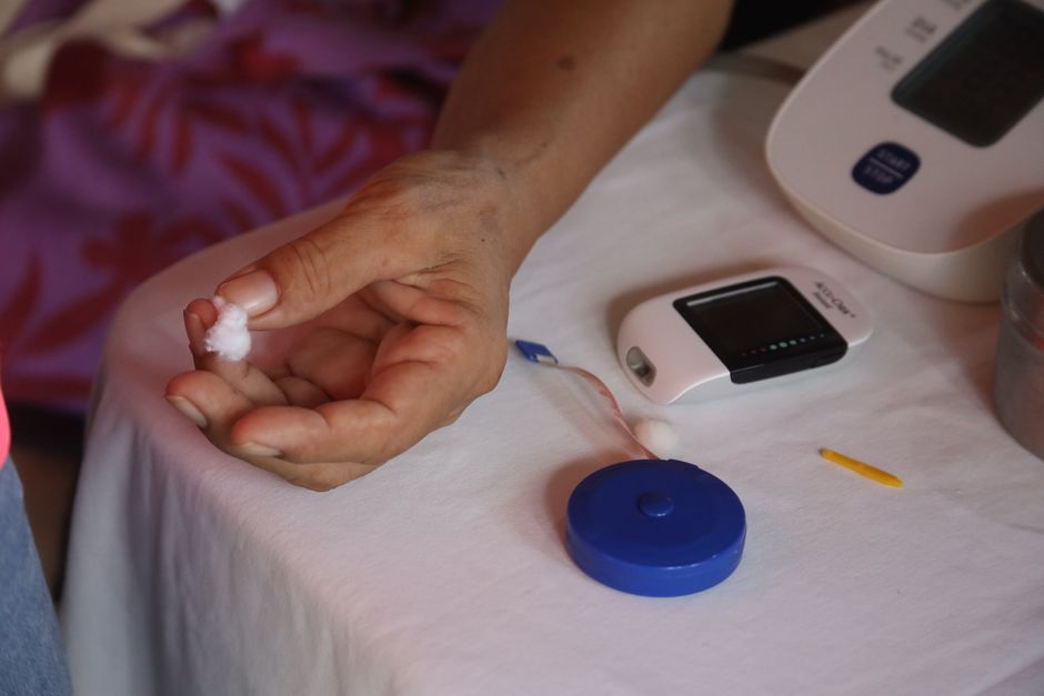
Cholestasis means bile flow has slowed or stopped. Bile is a digestive fluid made in the liver. When it cannot flow normally, bile salts and bilirubin build up in the blood. Testing for cholestasis relies primarily on blood tests that measure these substances. Imaging tests like ultrasound help find the cause. Together, these tools give doctors a clear picture of what is happening.
What Blood Tests Diagnose Cholestasis?
Blood tests are the first step. They look for signs that bile is backing up into the bloodstream. The key markers are bilirubin, alkaline phosphatase, and gamma-glutamyl transferase (GGT).
Bilirubin is a yellow waste product from broken-down red blood cells. The liver normally processes it and sends it into bile. When bile flow is blocked, bilirubin rises in the blood. High levels cause jaundice, the yellowing of skin and eyes.
Alkaline phosphatase (ALP) is an enzyme found in the bile ducts. When bile cannot flow, pressure builds and ALP leaks into the blood. Elevated ALP is a strong sign of cholestasis. However, ALP also comes from bone, so doctors often check GGT to confirm the source.
GGT is another enzyme concentrated in the liver and bile ducts. If ALP is high but GGT is normal, the ALP likely comes from bone. If both are elevated, the liver is the source. This combination is a standard way to confirm cholestasis on blood work.
What Do the Test Results Mean?
Normal ranges vary slightly by laboratory. In general, total bilirubin falls between 0.1 and 1.2 mg/dL. Alkaline phosphatase normal range is roughly 44 to 147 IU/L for adults. GGT normal range is about 8 to 61 IU/L. These are reference points, not strict rules. Your doctor interprets results in context.
In cholestasis, bilirubin may be elevated or normal. This depends on the type. In intrahepatic cholestasis, where the problem is inside the liver, bilirubin is often high. In some cases, ALP and GGT rise while bilirubin stays normal. This is called anicteric cholestasis. It means bile flow is impaired but jaundice has not developed.
Doctors also check liver enzymes ALT and AST. These measure liver cell damage. In cholestasis, they may be mildly elevated or normal. When they are very high, it suggests hepatitis or another liver injury rather than a pure bile flow problem.
How Do Imaging Tests Help Diagnose Cholestasis?
Blood tests confirm that cholestasis is present. Imaging tests find out why. The most common first imaging test is an abdominal ultrasound. It is painless, quick, and uses no radiation.
Ultrasound can show whether the bile ducts are dilated, or widened. Dilated ducts suggest a blockage. Gallstones are a frequent cause and show up clearly on ultrasound. The test also looks at the liver, pancreas, and gallbladder for masses or other abnormalities.
If ultrasound does not give a clear answer, doctors may order magnetic resonance cholangiopancreatography (MRCP). This is an MRI that creates detailed images of the bile ducts. It is non-invasive and very accurate at finding blockages, strictures, or stones that ultrasound missed.
Endoscopic retrograde cholangiopancreatography (ERCP) is both a diagnostic and treatment tool. A scope is passed through the mouth into the small intestine. Dye is injected into the bile ducts, and X-rays are taken. If a stone or stricture is found, it can often be treated during the same procedure. ERCP carries more risk than other imaging tests, so it is reserved for cases where intervention is likely needed.

What Causes Cholestasis?
Cholestasis has two broad categories. Intrahepatic cholestasis means the problem is inside the liver. Extrahepatic cholestasis means the problem is in the bile ducts outside the liver.
Common causes of intrahepatic cholestasis include drug-induced liver injury, viral hepatitis, primary biliary cholangitis, and alcoholic liver disease. Certain medications can impair bile flow. Some antibiotics, anabolic steroids, and oral contraceptives are known culprits.
Extrahepatic cholestasis is usually caused by a physical blockage. Gallstones are the most common cause. Tumors of the bile duct or pancreas can also block flow. Strictures, or narrowed areas, may develop after surgery or from chronic inflammation.
Pregnancy can trigger a specific form called intrahepatic cholestasis of pregnancy. It typically appears in the third trimester and causes severe itching. Blood tests show elevated bile acids. This condition requires monitoring because it carries risks for the baby.
When Should You See a Doctor?
See a doctor promptly if you notice jaundice. Yellowing of the skin or eyes is never normal. Dark urine and pale stools are also warning signs. These suggest bilirubin is building up in the blood and not reaching the intestines.
Severe itching without a rash is another symptom of cholestasis. Bile salts accumulate in the skin and cause intense itching. This is especially important in pregnancy, where it may indicate cholestasis of pregnancy.
Right upper abdominal pain can accompany cholestasis. This is the area where the liver and gallbladder sit. The pain may be dull and constant or sharp and intermittent. Fever with pain suggests an infection and requires urgent evaluation.
What Happens After Diagnosis?
Treatment depends on the cause. If a medication is responsible, stopping it often resolves the problem. Gallstones may require surgery to remove the gallbladder. Strictures can be treated with ERCP and stent placement.
For chronic conditions like primary biliary cholangitis, treatment focuses on slowing disease progression. Ursodeoxycholic acid is a medication commonly used for this purpose. It helps improve bile flow and liver enzyme levels in many patients.
Lifestyle changes can support liver health. Avoiding alcohol is important. Maintaining a healthy weight reduces the risk of fatty liver disease. Some doctors recommend a diet low in saturated fats, though dietary changes alone do not cure cholestasis.
The outlook depends entirely on the underlying cause. Acute causes like gallstones or drug reactions often resolve completely with treatment. Chronic causes may require long-term management. Regular follow-up with blood tests is essential to monitor liver function.
Can Cholestasis Be Prevented?
Not all types of cholestasis can be prevented. Genetic conditions and autoimmune diseases are not avoidable. However, some risk factors are modifiable.
Limiting alcohol intake protects the liver. Avoiding unnecessary medications reduces the risk of drug-induced cholestasis. Maintaining a healthy weight lowers the risk of gallstones. Rapid weight loss can actually increase gallstone risk, so gradual changes are better.
If you have liver disease, staying current with medical care is the best prevention strategy. Regular blood tests can catch problems early, before symptoms develop. Early detection improves outcomes in most liver conditions.
Frequently Asked Questions
What is the most accurate test for cholestasis?
Blood tests measuring bilirubin, alkaline phosphatase, and GGT are the most accurate for confirming cholestasis. Imaging tests like MRCP are most accurate for finding the cause.
Can cholestasis be seen on an ultrasound?
Ultrasound can show dilated bile ducts, gallstones, and some liver abnormalities. It cannot diagnose cholestasis itself, which requires blood tests.
How long does it take to get cholestasis blood test results?
Most blood test results are available within 24 to 48 hours. Imaging results may take slightly longer depending on the facility and type of test.
Is fasting required for cholestasis blood tests?
Some doctors recommend fasting for 8 to 12 hours before liver function tests. Follow your doctor’s specific instructions for accuracy.
