
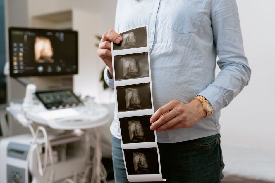
Ultrasound imaging uses high-frequency sound waves to create pictures of the inside of the body. A device called a transducer sends sound waves into the body and then listens for the echoes that bounce back. A computer turns those echoes into real-time images that show organs, tissues, and blood flow without using any radiation.
How Does Ultrasound Imaging Work Step by Step?
Sound waves travel through the body until they hit a boundary between different types of tissue. Some waves bounce back. The rest continue deeper until they hit another boundary.
The transducer is the key. It works as both a speaker and a microphone. It sends out short pulses of sound — far above the range of human hearing — and then switches to listening mode. Those returning echoes carry information about how deep each boundary is and how dense the tissue is.
Here is the basic sequence:
- The transducer sends a pulse of high-frequency sound into the body.
- Sound waves travel through tissue and reflect at boundaries between different densities.
- The transducer detects the returning echoes.
- A computer measures how long each echo took to return and how strong it is.
- The system converts that data into a two-dimensional image on a screen.
The physics behind this is straightforward. Sound travels through soft tissue at roughly 1,540 meters per second. That speed is fairly consistent across most soft tissues, which is what allows the machine to estimate depth from echo timing. When sound hits a boundary between tissues of different density — say, between muscle and fat — some of it reflects. The bigger the difference in density, the stronger the echo.
Different tissues reflect sound differently. Fluid like urine or amniotic fluid appears dark (called anechoic) because sound passes through it with almost no reflection. Dense tissue like bone reflects almost all sound and appears bright. This is why a bladder full of fluid looks black on an ultrasound screen while a kidney stone looks bright white.
What Is the Difference Between Ultrasound and Other Imaging Tests?
Ultrasound is one of several imaging tools, and each has distinct strengths and limitations. The main difference is that ultrasound uses sound waves while X-rays and CT scans use ionizing radiation.
| Imaging Type | Energy Source | Best For | Radiation? |
|---|---|---|---|
| Ultrasound | High-frequency sound waves | Soft tissue, organs, blood flow, pregnancy | No |
| X-ray | Ionizing radiation | Bones, lungs, dental | Yes |
| CT scan | Ionizing radiation | Detailed cross-sections of many body areas | Yes |
| MRI | Magnetic fields and radio waves | Brain, spinal cord, joints, soft tissue | No |
Ultrasound has real advantages. It is portable, relatively low-cost, and does not use ionizing radiation. It shows movement in real time, which makes it useful for watching a heart beat or guiding a needle during a procedure. MRI also avoids radiation but is more expensive and slower, and it cannot be used for real-time guidance in the same way.
Ultrasound has limits too. Sound waves do not travel well through bone or air. That means ultrasound cannot image structures behind bone, like the brain in adults, or areas filled with gas, like much of the bowel. Image quality also depends heavily on the skill of the person holding the transducer.
What Is Ultrasound Imaging Used For?
Ultrasound is used across many areas of medicine. Its ability to show soft tissue and fluid in real time makes it useful in situations where other imaging methods fall short.
Common uses include:
- Pregnancy — checking fetal growth, position, and anatomy
- Abdominal organs — liver, gallbladder, kidneys, pancreas, spleen
- Heart — evaluating heart valves, chambers, and pumping function (echocardiography)
- Blood vessels — detecting clots, narrowing, or abnormal blood flow
- Muscles and joints — tendons, ligaments, and fluid buildup
- Thyroid and breast — evaluating lumps or nodules
- Guiding procedures — helping doctors place needles or catheters accurately
One of the less obvious uses is guiding medical procedures. Because ultrasound shows real-time images, a doctor can watch a needle move through tissue on a screen and steer it to the right spot. This is common for biopsies, nerve blocks, and draining fluid collections.
Doppler ultrasound is a related technique that measures blood flow. It works on the same principle as a police radar gun — sound waves bouncing off moving blood cells shift in frequency. That shift tells the machine how fast and in what direction blood is flowing. It is used to check for narrowed arteries, blood clots, and problems with heart valves.





Is Ultrasound Safe?
Diagnostic ultrasound has been used in medicine for decades and is generally considered safe when used by trained professionals for standard imaging. It does not use ionizing radiation, which is the main concern with X-rays and CT scans.
That said, ultrasound is not completely without biological effects. Sound waves carry energy, and at high intensities that energy can heat tissue slightly. Diagnostic ultrasound uses low intensities and short exposure times, and current evidence does not show harm from standard diagnostic use. But the principle of “as low as reasonably achievable” still applies — the goal is to get the information needed with the shortest exposure and lowest power that will produce a useful image.
This matters most in pregnancy. Ultrasound is the most common imaging test used during pregnancy, and its safety record is well established for standard diagnostic exams. However, organizations that set ultrasound safety guidelines recommend that ultrasound during pregnancy be performed only when there is a medical need, by trained personnel, and that keepsake or entertainment ultrasound sessions without a medical purpose are not recommended.
No clinical guidelines currently support the use of prenatal ultrasound for non-medical purposes like 3D keepsake images. The concern is not that a single brief exposure has been shown to cause harm, but that the effects of prolonged or repeated exposure without medical justification have not been well studied.
What Does an Ultrasound Appointment Feel Like?
A standard ultrasound exam is painless. You lie on an exam table, and a technician applies a clear gel to your skin. The gel eliminates the air gap between the transducer and your skin, because air blocks sound waves.
The technician presses the transducer against your skin and moves it around to capture images from different angles. You may feel mild pressure, especially if the area being examined is tender. For some exams, you may need to hold your breath briefly or change position.
Preparation depends on the type of exam. Some abdominal ultrasounds require you to fast for several hours beforehand so that gas and food do not block the view of organs. Pelvic ultrasounds may require a full bladder, which pushes other structures out of the way and gives a clearer window. Others, like a thyroid or breast ultrasound, need no preparation at all.
Most exams take between 15 and 45 minutes, depending on the area being studied. Results are usually reviewed by a radiologist or your doctor, who interprets the images and sends a report.
What Are the Limitations of Ultrasound Imaging?
Ultrasound cannot see through everything. Bone blocks sound waves almost completely, which is why ultrasound is not used to image the brain in adults or to look at structures behind the ribs without special techniques. Air is another barrier — gas in the stomach or intestines can obscure the organs behind it.
Body size also affects image quality. Sound waves weaken as they travel through tissue, so imaging deeper structures in larger patients can be more difficult. Technologists adjust frequency and other settings to compensate, but there are physical limits.
Operator skill is a significant factor. Unlike a CT scan, which produces standardized images that any radiologist can read, ultrasound images depend on the person holding the probe. Two technicians can get different images of the same patient. This is not a flaw in the technology itself — it is a characteristic of how it works.
Ultrasound also cannot always tell the difference between benign and malignant findings on its own. A suspicious mass seen on ultrasound usually needs a biopsy to determine whether it is cancer. Ultrasound can guide that biopsy, but it cannot replace tissue analysis.
How Has Ultrasound Technology Changed?
Ultrasound has improved significantly since it was first used in medicine in the 1950s. Early machines produced static, grainy images. Modern systems produce high-resolution real-time video and can create 3D and 4D images.
Portable ultrasound devices have changed how and where ultrasound is used. Handheld units can now be carried to a patient’s bedside, used in ambulances, or taken to rural clinics. This has expanded access to imaging in settings where a full radiology suite is not available.
Contrast-enhanced ultrasound is another development. It uses tiny gas-filled microbubbles injected into the bloodstream to improve visibility of blood flow in organs like the liver. This technique is used in some countries more than others, and its availability varies.
Elastography is a newer method that measures how stiff or soft tissue is. Since some cancers and liver disease make tissue stiffer, this can add useful information. It is an active area of research, and its role in routine clinical practice continues to evolve.
Frequently Asked Questions
Does ultrasound use radiation?
No. Ultrasound uses high-frequency sound waves, not ionizing radiation. This is one reason it is commonly used during pregnancy and in children.
How long does an ultrasound take?
Most ultrasound exams take between 15 and 45 minutes. The time depends on the area being examined and how many images the doctor needs.
Can ultrasound detect cancer?
Ultrasound can show masses and other abnormalities, but it cannot confirm whether something is cancer. A biopsy is usually needed to make that diagnosis.
Why can’t ultrasound see through bone?
Bone is much denser than soft tissue and reflects almost all sound waves. This prevents the sound from reaching structures behind the bone.
