
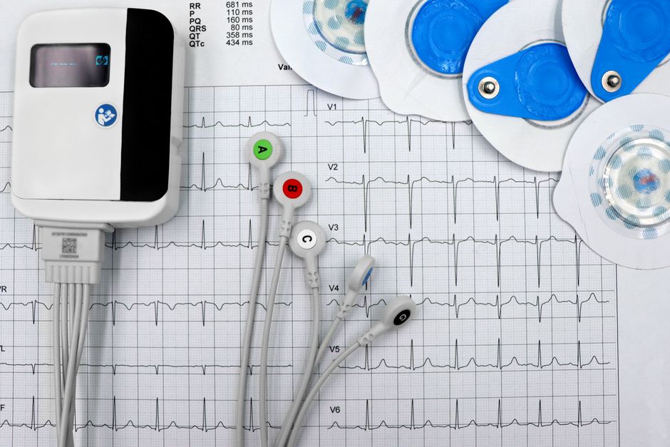
A focal seizure starts in one specific area of the brain. It does not begin everywhere at once the way a generalized seizure does. Because of that, diagnosing one is less like flipping a single switch and more like narrowing down a location. Doctors use a combination of tools — a detailed history, an electroencephalogram (EEG), brain imaging such as MRI, and sometimes blood tests or genetic testing — to confirm what is happening, find where it starts, and rule out other causes.
No single test does all of this alone. The EEG shows how the brain’s electrical activity behaves. The MRI shows what the brain tissue looks like. Together with the patient’s own account of their symptoms, these pieces form the diagnosis. This article walks through each step, what it can and cannot tell you, and why the order of testing matters.
What Makes a Seizure “Focal”?
A focal seizure begins in a network of neurons limited to one region of one hemisphere of the brain. A generalized seizure, by contrast, involves both hemispheres from the start. This distinction is not just a label — it changes which tests are useful and which treatments are considered.
The brain is organized by function. Different regions handle movement, sensation, language, memory, and emotion. When abnormal electrical activity stays in one region, the symptoms reflect that region’s job. A seizure in the part of the brain controlling the right hand may cause twitching there. One in a region involved with smell might produce a strange odor that is not really there.
This is why focal seizures can look so different from person to person. The symptoms are a clue to location. Doctors call this the semiology of the seizure — the observable signs and the symptoms the person reports. Careful attention to semiology often points toward the region where the seizure starts before any test is ordered.
Focal seizures are sometimes described as having “awareness” or “impaired awareness.” This replaced older terms like “simple partial” and “complex partial.” A person with impaired awareness may stare, appear confused, or make repetitive movements and not remember the event afterward. These details matter for diagnosis and for safety.
Why the Medical History Comes First
The most important diagnostic tool is not a machine. It is a careful conversation. Before any EEG or scan, a clinician wants to know exactly what happened, what the person felt, how long it lasted, and what they remember.
Witnesses are valuable here. A person having a focal seizure with impaired awareness often cannot describe it accurately. Someone who saw it happen can describe staring, lip-smacking, fumbling movements, or confusion that the person themselves may not recall.
Several conditions can mimic focal seizures, and the history helps separate them. These include fainting (syncope), migraine with unusual symptoms, transient ischemic attacks (sometimes called mini-strokes), sleep disorders, and certain movement disorders. Each has features that differ from a seizure, and the pattern of events often makes the difference clear.
Timing matters too. Seizures that happen shortly after waking, or that cluster around a specific trigger, give useful clues. So does the person’s age when symptoms began, any family history of seizures, any prior head injury or infection affecting the brain, and any medications being taken.
This history-taking is not a formality before the “real” tests. It frequently shapes which tests are done and how the results are interpreted.
What Does an EEG Actually Show?
An EEG records the brain’s electrical activity using small electrodes placed on the scalp. It is the primary test for detecting the abnormal electrical patterns associated with seizures.
The challenge is that a routine EEG is a snapshot. It typically lasts somewhere around 20 to 30 minutes. Many people with epilepsy have normal brain activity between seizures, so a routine EEG can come back normal even when seizures are real. A normal EEG does not rule out epilepsy.
To improve the odds of catching something useful, clinicians use several strategies:
- Sleep deprivation before the test, which can make abnormal patterns more likely to appear.
- Activation methods such as rapid breathing (hyperventilation) or flashing lights, which can trigger abnormal activity in some people.
- Longer recordings — an EEG that runs for several hours, overnight, or several days. Video EEG monitoring, done in a hospital or specialized unit, records both brain activity and the person on camera. This lets clinicians match what the brain is doing to what the seizure looks like.
Video EEG monitoring is often the most informative test when the diagnosis is unclear. If a person has an event while being monitored, clinicians can see whether it is truly a seizure and, if so, where in the brain it starts.
EEG findings can also be normal in a person who does have epilepsy, and abnormal in some people who do not have seizures. This is why the EEG is read alongside the history, not in isolation. The pattern of electrical activity, the person’s symptoms, and the imaging all inform one another.





What Is an MRI Looking For?
An MRI (magnetic resonance imaging) produces detailed pictures of the brain’s structure. Where the EEG shows function, the MRI shows anatomy. Both matter.
For someone with focal seizures, the MRI is looking for a structural cause — something in the brain tissue that could explain why seizures start where they do. Possible findings include:
- Scarring from a prior injury, stroke, or infection.
- Tumors, including slow-growing ones that may have been present for years.
- Malformations of cortical development — areas where the brain’s outer layer formed abnormally before birth.
- Hippocampal sclerosis, a pattern of scarring in a deep brain structure called the hippocampus, which is a common finding in temporal lobe epilepsy.
- Vascular malformations, which are abnormal tangles of blood vessels.
Not every MRI shows a clear cause. In a meaningful share of people with focal epilepsy, the MRI appears normal. This is called MRI-negative epilepsy. It does not mean the seizures are not real or that nothing is wrong — it means the cause is not visible on standard imaging.
When a standard MRI is normal but the diagnosis remains unclear, doctors may order a specialized epilepsy protocol MRI. This uses thinner image slices and specific sequences designed to detect subtle abnormalities that a routine scan can miss. The difference between a general MRI and an epilepsy-specific one can be significant.
MRI is generally considered safe, but it uses a strong magnetic field. People with certain implanted devices, such as some pacemakers or metal fragments in the eye, may not be able to have one. This is screened before the scan.
How EEG and MRI Work Together
Neither test is complete on its own. The EEG tells you that the brain’s electrical activity is abnormal and roughly where. The MRI tells you whether there is a physical reason for it. When the two agree — an EEG pattern pointing to the left temporal region and an MRI showing scarring in that same area — the diagnosis becomes much more confident.
When they disagree, or when both are normal, the picture is murkier. This is where longer monitoring, specialized imaging, and sometimes additional testing come in. The goal is to localize the seizure onset zone as precisely as possible, because that information guides treatment decisions.
What Other Tests Might Be Used?
EEG and MRI are the core tools, but they are not always the whole story. Depending on the situation, other tests may be ordered.
Blood tests can check for imbalances — such as abnormal sodium, calcium, or glucose levels — that can provoke seizures. They can also screen for infections or other conditions. Blood tests are more about ruling out reversible causes than confirming epilepsy itself.
Genetic testing is increasingly used, especially when seizures begin in childhood, when there is a family history, or when other features suggest an inherited condition. Many epilepsy-related genes have been identified, though the genetics of epilepsy remain complex and not every case has a known genetic cause.
Lumbar puncture (spinal tap) is occasionally used when an infection or inflammatory condition affecting the brain or its covering is suspected. It is not a routine part of focal seizure diagnosis.
Neuropsychological testing can assess memory, attention, and other cognitive functions. This is sometimes helpful when seizures involve areas important for thinking and memory, and it can inform treatment planning.
For people being evaluated for epilepsy surgery, additional specialized tests may be used, such as PET scans, SPECT scans, or intracranial EEG (electrodes placed directly on or in the brain). These are reserved for complex cases.
Why Getting the Diagnosis Right Matters
An accurate diagnosis changes everything that follows. It determines whether treatment is needed, which medication might be appropriate, and what safety precautions make sense. It also prevents the harm of treating something that is not a seizure — or missing something that is.
Some people are diagnosed with epilepsy who do not have it, and some people with epilepsy are told their episodes are something else. Both errors carry costs. That is why the process is deliberate: history first, then EEG, then imaging, then re-evaluation as needed.
Diagnosis is not always a single moment. For many people it is a process that unfolds over time as more information becomes available. That can feel frustrating, but it reflects the genuine difficulty of distinguishing between conditions that can look alike.
If you or someone you know is being evaluated for focal seizures, the most useful thing you can do is pay close attention to what happens during an episode and write it down. Note the time, what the person was doing, what they looked like, how long it lasted, and what they remember afterward. That record is often more valuable to a neurologist than any single test result.
Frequently Asked Questions
Can an EEG be normal if you have focal seizures?
Yes. A routine EEG is a short recording, and many people with epilepsy have normal brain activity between seizures. A normal EEG does not rule out epilepsy, which is why longer monitoring is sometimes needed.
What does an MRI show for focal seizures?
An MRI shows the structure of the brain and can reveal causes such as scarring, tumors, or malformations that may explain where seizures start. In some people, the MRI appears normal even when seizures are confirmed.
How long does it take to diagnose focal seizures?
There is no fixed timeline. Some people are diagnosed after a single evaluation, while others need repeated testing or extended monitoring over weeks or months. The pace depends on how clear the findings are.
Do you need both an EEG and an MRI to diagnose focal seizures?
Both are commonly used because they provide different information — the EEG shows electrical activity and the MRI shows brain structure. The combination helps confirm the diagnosis and locate where seizures begin.
