
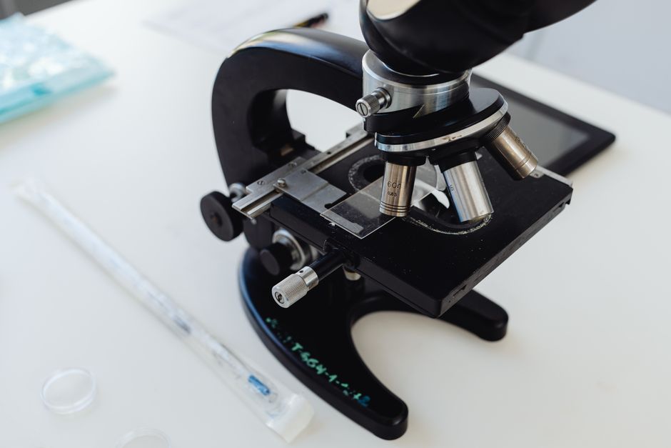
A compound microscope flips the image because of how its two lenses are arranged. Light from the specimen passes through the objective lens, which forms a real, inverted image inside the tube. The eyepiece then magnifies that already-flipped image, so what reaches your eye is upside down and reversed left to right. This is normal optics, not a defect.
That single fact explains most of the confusion people have when they first use a microscope. Move the slide right, and the specimen appears to move left. Nudge it toward you, and it seems to drift away. The instrument is working correctly. Your brain is the part that needs adjusting.
Why Is The Image Inverted In A Microscope?
Two lenses, two flips, and only one of them sticks.
A compound microscope uses an objective lens close to the specimen and an eyepiece lens near your eye. The objective is a converging lens with a very short focal length. When the specimen sits just beyond that focal point, the objective projects a real image into the body tube. Real images formed this way are inverted relative to the object.
That inverted image then acts as the object for the eyepiece. The eyepiece works like a simple magnifying glass. It takes the image already floating inside the tube and magnifies it without flipping it again. So the final image your eye receives is inverted once, not twice.
This is the part most people miss. A single magnifying glass does not invert anything. A telescope with two lenses can produce an upright image depending on the lens spacing. The compound microscope inverts because the objective forms a real image and the eyepiece simply enlarges it.
What Does Inverted Actually Mean Here?
Inverted means rotated 180 degrees, not just flipped top to bottom.
When the image is inverted, it is upside down and reversed left to right at the same time. Think of spinning a photograph half a turn on a table. The top goes to the bottom, and the left side goes to the right.
This matters because it changes how you move the slide. If you want to look at something near the top right of your field of view, you must move the slide toward the bottom left. The specimen and the image move in opposite directions.
Some people describe this as a mirror image. That is not quite right. A mirror flips front to back, not left to right. What a microscope does is a rotation, and the distinction becomes obvious the moment you try to trace a letter under the lens.
Why Does Moving The Slide Feel Backwards?
Your hand moves one way and the image moves the other because of the same 180-degree rotation.
The stage holds the specimen. The optics project an inverted version of it. When you push the slide to the right, the specimen moves right, but its projected image moves left across your field of view. Your visual system expects the world to move with your hand, and it does not here.
This is why new microscope users chase a specimen around the slide for the first few minutes. The correction is simple once you know it. Move the slide in the direction opposite to where you want the image to go.
With practice the reversal stops feeling wrong. Most experienced users stop thinking about it entirely and their hands adjust automatically.





Do All Microscopes Invert The Image?
No. It depends on the design of the instrument.
Simple microscopes, which are essentially a single magnifying lens, do not invert the image. Dissecting microscopes, also called stereo microscopes, use a different optical path with prisms or additional lenses that correct the orientation. That is why you can work on a specimen under a dissecting scope with your hands and tools without reversing your movements.
Standard compound microscopes used in classrooms and labs invert the image. So do most brightfield microscopes without added correction optics.
Some compound microscopes include an erecting prism or a similar optical element that flips the image back to upright. These are less common in basic teaching labs and more common in certain research or clinical setups. If you are unsure which type you have, place a letter “e” on a slide and look at it. If it appears upside down and backwards, your microscope inverts.
How Do You Work With An Inverted Image?
You learn to reverse your movements, and you learn to read the slide differently.
The practical adjustments are straightforward:
- Move the slide opposite to the direction you want the image to travel.
- When scanning for a structure, work in a consistent pattern so you do not lose your place.
- Remember that a specimen near the top of the field is actually near the bottom of the slide.
- Practice with a letter or a known shape until the reversal becomes automatic.
Reading a prepared slide takes a moment of translation. A tissue section that appears to run from upper left to lower right is actually running the other way on the glass. This matters when you are comparing a slide to a diagram or a textbook image.
Digital cameras and phone adapters change this experience. When you capture an image through the eyepiece, the camera records what the optics produce. Some software flips it back automatically. Some does not. If your captured image looks wrong compared to the slide, check the software settings before assuming the microscope is faulty.
Does The Magnification Change The Inversion?
No. Changing the objective does not change whether the image is inverted.
Switching from a 4x objective to a 40x objective changes how much the image is enlarged and how much detail you can see. It does not change the orientation. The image stays inverted at every magnification on a standard compound microscope.
What does change is your field of view. Higher magnification shows a smaller area. That makes the reversed movement feel more sensitive, because a small nudge of the slide shifts the image a large distance across a narrow field. This is a common source of frustration when people move from low to high power.
The fix is to center your specimen at low power first, then switch objectives. Centering is easier when the field is wide, and the specimen stays in view as you increase magnification.
Is This Related To How The Eye Works?
Yes, and the parallel is worth understanding.
The human eye also forms an inverted image on the retina. The lens of the eye focuses light onto the back of the eye, and the image there is upside down and reversed. Your brain interprets it as upright because it has learned to do so through years of visual experience.
A microscope adds a second inversion on top of the one your eye already handles. Your brain corrects for the eye’s inversion automatically. It does not correct for the microscope’s inversion, because that is a new and unnatural optical situation. That is why the reversed image feels wrong even though your eye is doing the same thing it always does.
People who wear glasses with prisms or who have used inverting lenses in experiments can adapt to a flipped world over time. The adaptation is real but slow, and it fades when the lenses come off. Microscope users get a smaller version of the same effect, which is why the reversal eventually stops bothering them.
Common Misconceptions About Microscope Images
A few ideas about microscope optics circulate widely and are worth correcting.
One is that the microscope is broken or assembled wrong when the image appears upside down. It is not. Inversion is the expected result of the standard two-lens design.
Another is that the eyepiece is responsible for the flip. The eyepiece magnifies. The objective is what forms the inverted real image.
A third is that higher magnification will somehow correct the orientation. It will not. Magnification and orientation are separate properties of the optical system.
Finally, some people assume all microscopes work the same way. They do not. Dissecting and stereo microscopes are designed to give an upright image, and that design choice is deliberate. It makes them usable for hands-on work where reversed movement would be a serious problem.
Frequently Asked Questions
Why is the image inverted in a microscope?
The objective lens forms a real, inverted image inside the tube, and the eyepiece magnifies that image without flipping it back. The result is a final image that is upside down and reversed left to right.
Do all microscopes show an inverted image?
No. Standard compound microscopes invert the image, but dissecting and stereo microscopes are designed to show it upright. Some compound microscopes also include correction optics that restore the upright orientation.
How do I move a slide when the image is upside down?
Move the slide in the direction opposite to where you want the image to go. If you want the image to move right, push the slide left.
Does higher magnification fix the inverted image?
No. Changing the objective changes how much the image is enlarged, not its orientation. The image stays inverted at every magnification on a standard compound microscope.
