
Blood gets oxygenated in the lungs, specifically in the alveoli — tiny air sacs where oxygen crosses into the blood and carbon dioxide leaves it. Deoxygenated blood arrives from the heart through the pulmonary arteries, picks up oxygen in the alveolar capillaries, and returns to the heart through the pulmonary veins. This exchange happens continuously with every breath, and it is the core purpose of the lungs.
Where Does Blood Get Oxygenated in the Lungs and Alveoli?
The actual transfer point is the alveolar-capillary membrane. This is a barrier so thin that oxygen and carbon dioxide can pass across it in a fraction of a second.
Here is the path in order:
- Deoxygenated blood enters the lungs through the pulmonary arteries, the only arteries in the body that carry oxygen-poor blood.
- These arteries branch into a dense network of capillaries that wrap around each alveolus.
- Oxygen from inhaled air moves across the alveolar wall into the blood. Carbon dioxide moves the other way, from blood into the air sac.
- Oxygen-rich blood collects into the pulmonary veins — the only veins carrying oxygen-rich blood — and returns to the left side of the heart.
- The heart pumps this blood out to the rest of the body through the aorta.
The wall between air and blood is extremely thin. That thinness is not a design flaw. It is what makes rapid gas exchange possible. If that membrane thickens or fills with fluid, oxygen transfer becomes harder, which is why many lung diseases cause low blood oxygen.
What Happens Inside the Alveoli During Gas Exchange?
Gas exchange in the alveoli works by simple diffusion. Molecules move from areas of higher concentration to areas of lower concentration, with no energy required from the body.
When you breathe in, the alveoli fill with air that is rich in oxygen. The blood arriving in the surrounding capillaries has just returned from delivering oxygen to your tissues, so its oxygen level is low. That difference in concentration is the driving force. Oxygen moves from the air into the blood. Carbon dioxide, which is higher in the blood than in the air you just inhaled, moves from the blood into the alveoli to be exhaled.
Diffusion is fast because the distance is tiny and the surface area is enormous. The lungs contain millions of alveoli. If you spread their combined surface flat, it would cover a large area — often compared to the size of a tennis court. That vast area, combined with the thin membrane, allows the blood to become fully oxygenated during the brief time it spends passing through the lung capillaries.
One detail worth knowing: the blood does not spend long in the capillaries. At rest, it passes through in a fraction of a second. During hard exercise, that time shortens because the heart is pumping faster. In healthy lungs there is still enough time to fully oxygenate the blood. In diseased lungs, that margin can disappear, which is one reason breathlessness on exertion is an early sign of lung problems.
How Do Oxygen and Carbon Dioxide Move in Opposite Directions?
They move in opposite directions because each gas follows its own concentration gradient. Oxygen is high in the air and low in the arriving blood, so it flows inward. Carbon dioxide is high in the blood and low in the air, so it flows outward.
These two gradients exist at the same time and at the same place. The membrane does not sort the gases. It simply lets both pass according to where each one is more concentrated. This is why the process is sometimes described as a two-way street.
Most carbon dioxide is not carried as a dissolved gas. The majority travels in the blood as bicarbonate, a form produced through a chemical reaction inside red blood cells. When that blood reaches the lungs, the reaction reverses and carbon dioxide is released into the alveoli to be breathed out. This matters clinically because the body’s acid-base balance is tied to how well the lungs remove carbon dioxide. If breathing becomes too slow or too shallow, carbon dioxide builds up, and that affects blood pH.




What Role Do Red Blood Cells and Hemoglobin Play?
Hemoglobin is the molecule inside red blood cells that actually carries oxygen. Without it, very little oxygen would dissolve in blood plasma, and the amount delivered to tissues would be far too low to sustain life.
Each red blood cell contains millions of hemoglobin molecules. Each hemoglobin molecule can bind up to four oxygen molecules. When blood passes through the alveolar capillaries, oxygen binds to hemoglobin, and the blood leaves the lungs nearly fully saturated. When that blood reaches tissues where oxygen is low, hemoglobin releases its oxygen.
This is a key point that is often misunderstood. The lungs do not “fill” the blood with oxygen the way you fill a tank. Instead, oxygen attaches to hemoglobin, and hemoglobin releases it later where it is needed. The system is a delivery service, not a storage tank.
Several things affect how tightly hemoglobin holds onto oxygen, including acidity, temperature, and carbon dioxide levels. In active tissues, these conditions shift so that hemoglobin releases more oxygen exactly where demand is highest. This is a normal and well-established part of how the body matches oxygen supply to demand.
What Can Prevent Blood From Getting Enough Oxygen?
Anything that disrupts the path from air to blood can lower blood oxygen. The problem can sit at any step: getting air into the lungs, moving it to the alveoli, crossing the membrane, or carrying oxygen in the blood.
Common categories include:
- Airway problems — conditions that block or narrow the airways, such as asthma or chronic obstructive pulmonary disease (COPD).
- Alveolar damage — diseases that destroy alveolar walls or fill the air sacs with fluid or inflammation, such as pneumonia or emphysema.
- Thickened or scarred membranes — conditions that make the alveolar wall less able to pass oxygen, such as pulmonary fibrosis.
- Reduced blood flow to the lungs — problems that limit how much blood reaches the exchange surface.
- Low hemoglobin or fewer red blood cells — anemia reduces the blood’s oxygen-carrying capacity even if the lungs work well.
These categories can overlap. A person can have more than one issue at once, which is why doctors use several tests rather than a single measurement.
How Do Doctors Measure Whether Blood Is Getting Oxygenated?
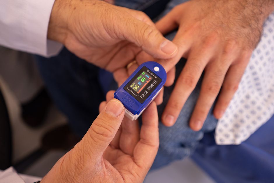
The most common measurement is pulse oximetry. A small device clips onto a finger and estimates oxygen saturation, which is the percentage of hemoglobin carrying oxygen. It is quick, painless, and widely used.
When a more precise measurement is needed, doctors use arterial blood gas testing. This involves drawing blood from an artery, usually in the wrist, and directly measuring oxygen, carbon dioxide, and acidity. It is more accurate than pulse oximetry but requires a needle draw.
Pulse oximetry has limits. It can be less reliable in poor circulation, with certain nail products, or with abnormal hemoglobin. When results seem inconsistent with how a person looks or feels, doctors often confirm with a blood draw.
Other tests look at the lungs themselves rather than the blood. Pulmonary function tests measure how much air you can move and how well it flows. Chest imaging can show structural problems in the alveoli or airways. Together, these tests help identify where in the oxygen pathway the problem lies.
Why Does This Matter for Everyday Health?
Understanding where blood gets oxygenated helps make sense of common symptoms. Shortness of breath, fatigue, and confusion can all point to problems with oxygen delivery, though each has many possible causes.
It also clarifies why certain habits matter. Smoking damages alveolar walls and airways, which reduces the surface area available for gas exchange. That damage is often permanent. Avoiding smoke exposure and managing conditions like asthma or COPD are the main ways to protect this system, based on long-standing clinical evidence.
One non-obvious point: oxygen saturation on a monitor does not tell you how well your tissues are using oxygen. It only reflects how much oxygen is riding on hemoglobin in that moment. A normal reading does not rule out every problem, and a low reading does not always mean the lungs are the cause. That is why doctors interpret these numbers alongside the full clinical picture rather than in isolation.
Frequently Asked Questions
Where exactly does blood get oxygenated?
Blood gets oxygenated in the alveoli, the tiny air sacs at the end of the airways in the lungs. Oxygen crosses from the air in the alveoli into the surrounding capillaries and binds to hemoglobin in red blood cells.
Does oxygen enter the blood in the lungs or the heart?
Oxygen enters the blood in the lungs, specifically in the alveolar capillaries. The heart pumps that oxygenated blood to the body but does not add oxygen to it.
What is the alveolar-capillary membrane?
It is the thin barrier between the air in the alveoli and the blood in the capillaries. Oxygen and carbon dioxide pass across it by diffusion, moving from higher to lower concentration.
Can low oxygen happen even if the lungs seem fine?
Yes. Anemia, heart problems, and other conditions can reduce oxygen delivery even when the lungs are working normally. This is why doctors look at the whole picture, not just one test.
