
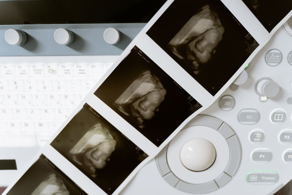
A fetal growth scan is typically done between 28 and 32 weeks of pregnancy. Some providers schedule it earlier, around 24 weeks, or later, up to 36 weeks, depending on your health and your baby’s development. This ultrasound measures your baby’s size and checks that growth is progressing as expected.
What Exactly Is a Fetal Growth Scan?
A fetal growth scan is a detailed ultrasound. It is different from the early dating scan or the anatomy scan done around 20 weeks. The purpose here is specific: to measure the baby and estimate weight.
The sonographer takes several measurements. These include the head circumference, abdominal circumference, and femur (thigh bone) length. These numbers are compared against standard growth charts for your baby’s gestational age.
The scan also checks the amount of amniotic fluid around the baby. It looks at the placenta’s position and health. Blood flow through the umbilical cord may also be assessed using Doppler ultrasound.
Why Is the Scan Done Between 28 and 32 Weeks?
This window is not random. It is the time when growth problems become detectable and when intervention can still make a difference.
Before 28 weeks, a baby that is small may simply be constitutionally small. After 32 weeks, the options for managing a growth problem become more limited. The 28-to-32-week window gives clinicians the best chance to identify issues and plan care.
Most pregnancies have one growth scan in this period. Some have more. The schedule depends entirely on your individual risk factors and what the first scan shows.
Who Needs a Growth Scan and When?
Routine scans. Many providers offer one growth scan to all pregnant patients around 30 weeks. This is standard practice in many clinics, even for low-risk pregnancies. It confirms the baby is growing along a normal curve.
High-risk pregnancies. If you have conditions like gestational diabetes, chronic high blood pressure, or preeclampsia, your doctor may order growth scans more often. These might start earlier and repeat every 2 to 4 weeks until delivery.
Previous complications. If you previously had a baby with growth restriction, your current pregnancy will be monitored more closely. The same applies if you have had a stillbirth or placental problems before.
Clinical concerns. If your fundal height — the measurement from your pubic bone to the top of your uterus — measures smaller or larger than expected, a growth scan is ordered. This is a common reason for an unscheduled scan.





What Happens If the Scan Shows a Problem?
First, understand that one measurement is not a diagnosis. Babies grow in spurts. A single scan showing a baby at the 15th percentile does not automatically mean there is a problem.
If the scan shows the baby is measuring small for gestational age, your provider will likely schedule a follow-up scan. This is usually done 2 to 3 weeks later to check the growth trajectory. A single low measurement is less concerning than a pattern of falling off the growth curve.
If the estimated fetal weight falls below the 10th percentile, the condition is called fetal growth restriction (FGR). This diagnosis triggers closer monitoring. Your doctor may order more frequent ultrasounds, non-stress tests, or Doppler studies of blood flow in the umbilical artery.
The key point is that detecting a problem early gives your medical team options. They can monitor more closely, adjust delivery timing, and make decisions based on data rather than guesswork.
Can You Have a Growth Scan Too Early or Too Late?
A growth scan done before 24 weeks is not reliable for assessing growth. The baby is too small, and normal variation is too wide. Scans done this early are more useful for dating the pregnancy than for measuring growth.
Scans done after 36 weeks have a different limitation. The baby is running out of room. Measurements become less accurate as the baby curls into the available space. The margin of error on weight estimates grows in the final weeks.
Most clinicians avoid relying on a single late scan for growth assessment. If late monitoring is needed, serial scans are preferred. This means repeating the scan at consistent intervals to track the trend rather than interpreting one snapshot.
How Accurate Is the Growth Scan?
Growth scans provide an estimate, not an exact measurement. The margin of error on estimated fetal weight is roughly plus or minus 15 percent. A baby estimated at 7 pounds could weigh anywhere from about 6 to 8 pounds at birth.
This variability is not a flaw in the technology. It is a limitation of measuring a moving baby through layers of tissue and fluid. Ultrasound measurements are mathematically derived from two-dimensional images.
The trend matters more than the absolute number. A baby whose growth follows a steady curve is generally doing well. A baby whose measurements stall or drop between scans warrants attention.
What Does the Scan Show About the Placenta?
The placenta is the baby’s lifeline. It supplies oxygen and nutrients. A growth scan evaluates the placenta’s position and, indirectly, its function.
Doppler ultrasound measures blood flow in the umbilical artery. High resistance in this vessel can indicate the placenta is not delivering blood efficiently. This finding supports a diagnosis of placental insufficiency.
Placental insufficiency is a leading cause of fetal growth restriction. It means the placenta is not keeping up with the baby’s demands. When this is detected, your care team will increase monitoring frequency.
Amniotic fluid levels are also part of the assessment. Low fluid can indicate the baby is not urinating enough, which may reflect reduced blood flow to the kidneys. It can also suggest a problem with the placenta.
Fetal Growth Scan vs. Anatomy Scan: What Is the Difference?
The anatomy scan, done around 18 to 22 weeks, looks for structural abnormalities. It examines the baby’s organs, limbs, spine, and heart. Its purpose is to detect birth defects.
The growth scan is a different exam with a different goal. It focuses on measurements and estimated weight. It does not examine fetal anatomy in detail.
Some women assume the 20-week anatomy scan covers everything. It does not assess growth adequacy. A baby can have a normal anatomy scan at 20 weeks and still develop growth restriction later in pregnancy.
Frequently Asked Questions
Is a fetal growth scan done at every pregnancy ultrasound?
No. A growth scan is a specific type of ultrasound with specific measurements. Early dating scans and anatomy scans serve different purposes and do not include growth assessment.
How long does a fetal growth scan take?
The scan typically takes 20 to 30 minutes. It may take longer if the baby is in a position that makes measurements difficult.
Do I need a full bladder for a growth scan?
Usually not. A full bladder is needed for early pregnancy scans. For a growth scan in the third trimester, most providers ask you to empty your bladder before the exam.
Can a growth scan tell the exact birth weight?
No. The scan provides an estimated fetal weight with a margin of error of about 15 percent. Only delivery reveals the exact birth weight.
