
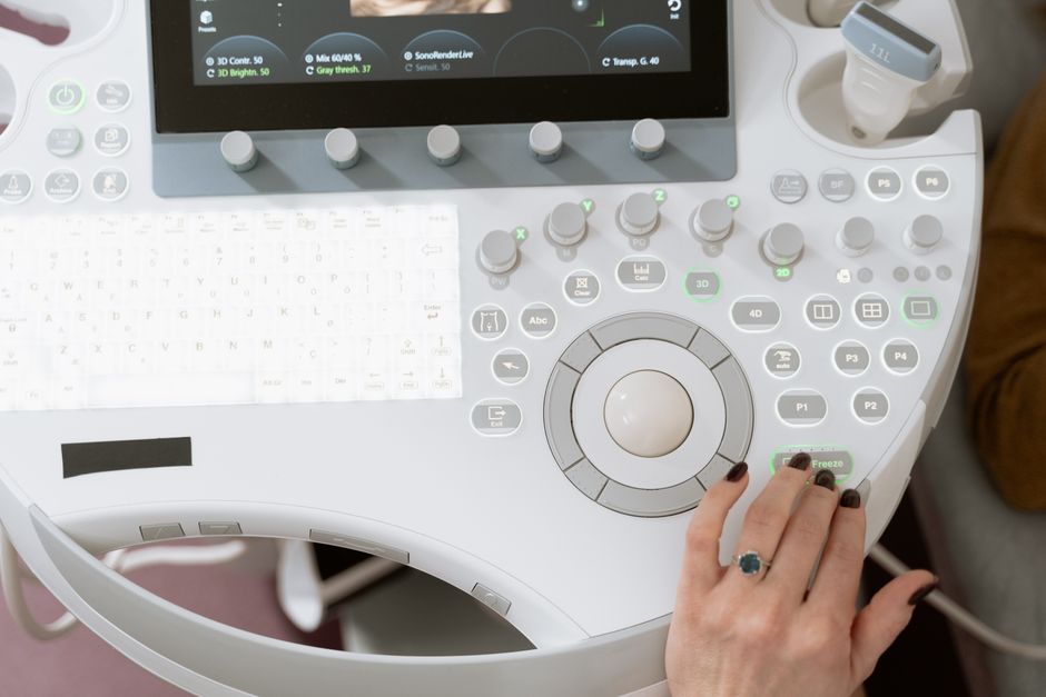
Pelvis sonography is a medical imaging test that uses high-frequency sound waves to create pictures of the organs and structures inside the pelvis. A small handheld device called a transducer sends sound waves into the body, and a computer turns the returning echoes into images. It uses no radiation, and it is one of the most common imaging tests ordered for pelvic symptoms.
The exam is also called a pelvic ultrasound. It can look at the uterus, ovaries, fallopian tubes, cervix, and the space around them in women. In men, it can look at the bladder, prostate, and seminal vesicles. It can also check blood flow, fluid collections, and lymph nodes in the pelvic area.
What Is Pelvis Sonography Used For?
Doctors order pelvic sonography for a wide range of reasons, and the list differs between men and women.
In women, it is often the first imaging test for pelvic pain, unusual bleeding, or a suspected problem with the uterus or ovaries. It can detect uterine fibroids, ovarian cysts, and thickened uterine lining. It helps evaluate possible causes of infertility and can confirm an early pregnancy and check its location. It is also used to guide a needle during certain procedures, such as draining a cyst or sampling tissue.
In men, pelvic sonography is used less often than in women, but it has clear roles. It can measure the prostate, check the bladder, and look for fluid buildup or cysts. A related form, transrectal ultrasound, is sometimes used to guide a prostate biopsy.
Across both groups, the test can answer questions such as:
- Is there a mass, cyst, or fluid collection?
- Is an organ enlarged or abnormal in shape?
- Is blood flowing normally through pelvic vessels?
- Has a pregnancy implanted in the right place?
One point worth knowing: ultrasound is very good at showing structure and fluid, but it cannot see through bone or air. That limits how much it can tell you about some deep pelvic structures, and it is one reason other imaging tests are sometimes needed.
How Does Pelvis Sonography Work?
Sound waves travel into the body and bounce back off tissue boundaries. Dense tissue, like muscle and bone, reflects more sound than fluid does. The machine reads these differences and builds a picture.
The frequency of the sound waves matters. Higher frequencies produce sharper images but do not travel as deep. Lower frequencies reach deeper structures but with less detail. The technologist chooses settings based on what needs to be seen and how much tissue the sound must pass through.
There are three main ways a pelvic ultrasound is done:
- Transabdominal: The transducer moves across the lower belly. You need a full bladder for this, because a full bladder pushes bowel out of the way and gives a clear window to see through.
- Transvaginal: A slim transducer is placed inside the vagina. It gives a closer, sharper view of the uterus and ovaries and does not require a full bladder.
- Transrectal: A transducer is placed in the rectum, mainly used in men to look at the prostate.
Doppler ultrasound is a related technique that measures blood flow. It can show whether a structure has a normal blood supply, which helps distinguish some types of masses from others.
What Happens During a Pelvic Ultrasound?
The test is usually quick and does not require sedation or recovery time. Most pelvic ultrasounds take between 15 and 45 minutes, depending on what is being checked and which approach is used.
For a transabdominal scan, you will be asked to drink water and hold your bladder before the appointment. The technologist applies a clear gel to your lower belly and moves the transducer across the skin. You may feel mild pressure but not pain.
For a transvaginal scan, you empty your bladder first. The transducer is covered with a sheath and lubricant, then gently inserted. Most people describe it as uncomfortable rather than painful. You can ask to stop at any point.
After the scan, a doctor trained in imaging reads the pictures and sends a report to the doctor who ordered the test. Results are usually available within a few days, though urgent findings are communicated sooner.





How Does Pelvis Sonography Compare to Other Imaging Tests?
Each imaging method has strengths and limits, and the right choice depends on the question being asked.
| Test | Best For | Radiation | Main Limits |
|---|---|---|---|
| Ultrasound | Uterus, ovaries, bladder, fluid, blood flow | None | Limited by bone and air; operator-dependent |
| CT scan | Bone, bowel, widespread disease, trauma | Yes | Ionizing radiation exposure |
| MRI | Soft tissue detail, staging, deep structures | None | Slower, costlier, not for some implants |
Ultrasound is often the first step because it is safe, fast, and does not use radiation. If more detail is needed, a doctor may order CT or MRI afterward. Ultrasound is not a replacement for these tests, and in some cases it simply cannot provide the information needed.
Is Pelvis Sonography Safe?
Diagnostic ultrasound has been used for decades, and no harmful effects have been confirmed at the energy levels used for imaging. It does not use ionizing radiation, which is its main advantage over CT.
That said, ultrasound is not without limitations. It is highly dependent on the skill of the person performing it and on the body habitus of the patient. Excess body fat, bowel gas, or scarring can reduce image quality. A normal ultrasound does not always rule out every condition, and a suspicious finding on ultrasound often needs confirmation with another test.
There is one area where caution is applied more carefully: pregnancy. Ultrasound is routinely used in pregnancy and is considered the standard imaging method when a scan is needed. However, the general principle in medicine is that imaging should be done when there is a clear medical reason, not for keepsake or non-medical purposes. This is a matter of professional guidance rather than evidence of harm at diagnostic levels.
What Can Pelvis Sonography Detect — and What Can It Miss?
Ultrasound is good at detecting fluid-filled structures, solid masses above a certain size, and changes in organ shape or size. It can show ovarian cysts, fibroids, fluid in the pelvis, and an enlarged prostate. Doppler can show abnormal blood flow.
It is less reliable for very small lesions, for structures hidden behind bowel gas, and for tissue that looks similar in density. It also cannot always tell whether a mass is benign or malignant. That distinction often requires a biopsy and lab analysis, not imaging alone.
This is an important point for patients: an ultrasound result is one piece of information. It is interpreted alongside your symptoms, your history, and sometimes other tests. A normal result does not automatically mean nothing is wrong, and an abnormal result does not automatically mean something serious.
Who Should Consider Pelvis Sonography?
Pelvic ultrasound is not a routine screening test for most people. It is ordered when there is a specific symptom or question to answer.
Common reasons a doctor might order one include:
- Pelvic pain or pressure that does not have a clear cause
- Abnormal vaginal bleeding, especially after menopause
- A suspected ovarian cyst, fibroid, or other mass
- Difficulty urinating or blood in the urine
- Evaluation of infertility
- Confirming and dating an early pregnancy
- Monitoring a known condition over time
If you are unsure whether a pelvic ultrasound is right for your situation, that is a conversation to have with your doctor. The test itself is low-risk, but whether it will answer your specific question depends on what is being looked for.
Frequently Asked Questions
What is pelvis sonography in simple terms?
It is an imaging test that uses sound waves to create pictures of the organs inside your pelvis, such as the uterus, ovaries, bladder, and prostate. It does not use radiation and is usually painless.
Do you need a full bladder for a pelvic ultrasound?
Only for the transabdominal approach, where a full bladder helps push bowel aside and gives a clearer view. A transvaginal ultrasound requires you to empty your bladder first.
How long does a pelvic ultrasound take?
Most pelvic ultrasounds take between 15 and 45 minutes. The time depends on which approach is used and what the doctor needs to see.
Can a pelvic ultrasound detect ovarian cancer?
It can show an ovarian mass, but it cannot reliably tell whether a mass is cancer. Confirming cancer usually requires a biopsy and lab testing, not imaging alone.
