
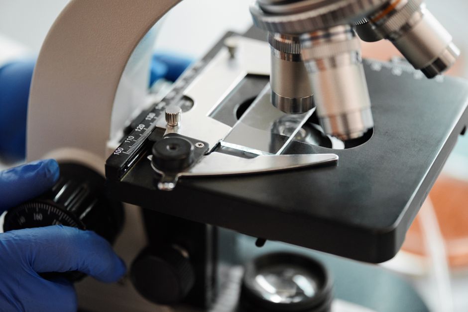
Immunofluorescence microscopy is a laboratory technique that uses antibodies tagged with fluorescent dyes to locate specific proteins, pathogens, or other target molecules inside cells and tissues. When those tagged antibodies bind to their targets, a microscope equipped with special filters and a light source makes the glow visible. That glow tells researchers and pathologists exactly where a target molecule sits, and sometimes how much of it is present.
The method has been a workhorse of biology and medicine since the mid-20th century. It shows up in hospital pathology labs, research universities, and drug development. What follows explains how it works, the main variations, where it is used, and where its limits lie.
What Is Immunofluorescence Microscopy and How Does It Work?
The technique rests on two well-established pieces of biology: the immune system’s ability to make antibodies that bind one specific target, and the ability of certain chemical dyes to absorb light at one wavelength and emit it at another.
Here is the basic sequence. A scientist takes an antibody that recognizes a chosen target — say, a particular protein on a cell surface. That antibody is chemically linked to a fluorescent molecule, called a fluorophore. The sample, which might be a slice of tissue or a layer of cultured cells, is treated with this labeled antibody. After washing away anything that did not bind, the sample goes under a fluorescence microscope.
The microscope shines light of one wavelength onto the sample. Wherever the labeled antibody has attached, the fluorophore absorbs that light and re-emits it at a longer wavelength. Optical filters separate the emitted light from the incoming light so only the glow is seen. The result is a bright signal against a dark background, pinpointing the target.
This is the core principle behind the direct and indirect methods described below. The specificity comes from the antibody. The visibility comes from the fluorophore.
What Is Immunofluorescence Microscopy Methods Uses?
There are two main methods, and they differ in how the fluorescent tag reaches the target.
In direct immunofluorescence, the antibody that recognizes the target is itself labeled with the fluorophore. This is a one-step process. It is fast and produces less background noise, but each target needs its own labeled antibody, and the signal from a single fluorophore can be faint.
In indirect immunofluorescence, an unlabeled primary antibody binds the target first. Then a second, labeled antibody that recognizes the primary antibody is added. Because several labeled secondary antibodies can attach to a single primary antibody, the signal is amplified. This makes indirect methods more sensitive and more flexible, since one labeled secondary antibody can work with many different primary antibodies. The trade-off is an extra step and slightly more background.
Both methods can be applied to cells grown in a dish or to thin slices of tissue. When the goal is to see the natural distribution of a molecule across a tissue section, the technique is often called immunofluorescence histochemistry. When it is applied to individual cells, it is called immunocytochemistry.
There is also a practical distinction based on what happens to the cells. In fixed samples, cells are chemically preserved so their structure is locked in place. This is the standard approach for most diagnostic and research work. In live-cell imaging, fluorescent proteins are often used instead of antibodies, because antibodies generally cannot cross an intact cell membrane.
What Is Immunofluorescence Used For in Medicine?
The technique has several established clinical and research roles.
In diagnostic pathology, it is used to detect the buildup of antibodies and complement proteins in tissue. This is central to diagnosing certain kidney diseases, where immune deposits along the filtering structures of the kidney produce characteristic patterns. It is also used in skin pathology to help identify autoimmune blistering conditions, where antibodies collect at specific layers of the skin.
In microbiology and infectious disease, immunofluorescence can detect specific bacteria, viruses, or other organisms in patient samples. Because it identifies organisms by their molecular markers, it can distinguish closely related species that look similar under a standard microscope.
In research, it is used constantly to answer questions about where proteins live inside cells, how they move, and how their location changes under different conditions. It also plays a role in drug development, where researchers check whether a treatment changes the distribution of a target molecule.
One non-obvious point: immunofluorescence shows location and presence, but it is not typically a precise way to measure how much of a molecule exists. For quantity, labs usually turn to other methods. The strength of immunofluorescence is spatial — telling you where something is.





What Is the Difference Between Direct and Indirect Immunofluorescence?
The table below lays out the practical differences between the two main approaches.
| Feature | Direct Immunofluorescence | Indirect Immunofluorescence |
|---|---|---|
| Steps | One labeled antibody | Primary plus labeled secondary |
| Signal strength | Lower | Higher, due to amplification |
| Flexibility | Needs a labeled antibody per target | One labeled secondary serves many targets |
| Background | Generally lower | Can be higher |
| Common use | Direct detection in tissue | Research and many diagnostic tests |
Neither method is better in every situation. The choice depends on how much signal is needed, how many targets are being studied, and whether background noise is a problem.
What Are the Limitations of Immunofluorescence Microscopy?
The technique is powerful, but it has real constraints, and knowing them matters for interpreting results.
Antibody specificity is the biggest issue. An antibody is supposed to bind one target, but some bind to unintended molecules. If that happens, the glow appears in the wrong place, and a researcher could draw a false conclusion. This is why careful controls are essential, and why results from a single antibody are usually confirmed with a second one.
Fluorescence fades. The dyes lose their brightness over time when exposed to light, a process called photobleaching. This limits how long a sample can be viewed and how many images can be captured.
Sample preparation can change what you see. Fixing and processing tissue can alter or hide some target molecules, so a negative result does not always mean the molecule is absent.
Background signal is a constant concern. Unbound antibodies and natural tissue fluorescence can both create glow that is not from the target. Good technique and proper controls reduce this, but it cannot be eliminated entirely.
Finally, standard immunofluorescence is not a reliable way to measure exact quantities. It shows presence and location well. It does not give a precise count of molecules.
How Does Immunofluorescence Compare With Other Microscopy Methods?
It helps to place the technique next to the alternatives.
- Standard light microscopy shows cell and tissue structure using stains, but it cannot identify specific proteins by name.
- Immunohistochemistry uses antibodies too, but the signal is a colored stain rather than a glow. It is common in clinical labs because it needs only a standard microscope and the results are permanent, not fading.
- Fluorescent proteins, such as those used in live-cell imaging, are built into the cell by genetic engineering rather than added by antibody. This allows living cells to be watched over time.
- Flow cytometry also uses fluorescently labeled antibodies, but it measures cells one at a time in a fluid stream rather than viewing them in place. It is better for counting large numbers of cells, while immunofluorescence is better for seeing location within tissue.
The common thread is that immunofluorescence sits in a middle ground. It gives the spatial detail that flow cytometry loses, and the molecular specificity that plain light microscopy cannot provide. It does not replace the others. It answers a different question.
Frequently Asked Questions
What is immunofluorescence microscopy in simple terms?
It is a lab method that uses antibodies attached to glowing dyes to show exactly where a specific molecule sits in a cell or tissue. The glow, viewed under a special microscope, marks the target’s location.
What is the difference between direct and indirect immunofluorescence?
Direct immunofluorescence labels the target-binding antibody itself, while indirect immunofluorescence adds a second labeled antibody that amplifies the signal. Indirect methods are more sensitive but involve an extra step.
Is immunofluorescence used to diagnose diseases?
Yes. It is used in pathology to help diagnose certain kidney and skin conditions by detecting immune deposits in tissue, and in microbiology to identify specific organisms. It is one tool among several that clinicians use together.
Can immunofluorescence measure how much of a protein is present?
Not precisely. It shows presence and location well, but it is not a reliable way to measure exact quantities. Labs use other methods when they need accurate amounts.
