Medical imaging lets doctors see inside your body without making a single cut. It includes X-rays, CT scans, MRIs, ultrasounds, and nuclear medicine tests that create pictures of bones, organs, and tissues. These images help find diseases early, guide treatments, and monitor how well therapies are working. Safety depends on the type of imaging—some use radiation, others do not—and each has specific risks and benefits that your doctor should explain before any test.
What Are the Main Types of Medical Imaging?
There are five major categories of imaging used in modern healthcare. Each works differently and serves a distinct purpose.
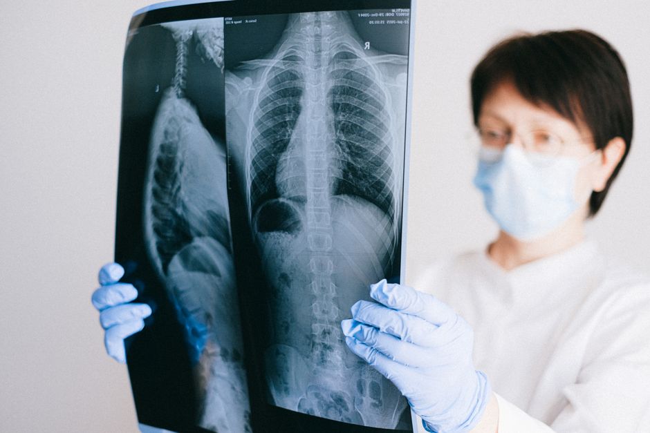
X-rays are the oldest and most common form. They use a small dose of ionizing radiation to create images of dense structures like bones. A chest X-ray can spot pneumonia or a collapsed lung. A dental X-ray finds cavities below the gum line. X-rays are quick, cheap, and widely available. The radiation dose is low but not zero.
CT scans (computed tomography) take X-ray technology further. The machine rotates around your body and combines multiple images into cross-sectional slices. CT scans provide much more detail than a standard X-ray. They are excellent for finding tumors, internal bleeding, and complex fractures. The radiation dose is higher than a plain X-ray—sometimes 100 to 200 times higher depending on the body part being scanned.
MRI (magnetic resonance imaging) uses strong magnets and radio waves, not radiation. It produces incredibly detailed images of soft tissues like the brain, spinal cord, muscles, and ligaments. MRIs are the gold standard for diagnosing torn ligaments, herniated discs, and certain types of brain tumors. The trade-off is time. A typical MRI takes 30 to 60 minutes and requires you to lie completely still inside a narrow tube.
Ultrasound uses high-frequency sound waves to create real-time images. It is the safest major imaging method because it involves no radiation. Ultrasounds are best for viewing soft tissues and fluid-filled structures. They are used routinely in pregnancy to check on fetal development. They also help diagnose gallbladder disease, thyroid nodules, and blood flow problems in veins and arteries.
Nuclear medicine and PET scans work differently from the others. You receive a small amount of radioactive material, either by injection or swallowing. Special cameras then detect where this material collects in your body. These scans show how organs and tissues are functioning, not just their structure. PET scans are commonly used to detect cancer, monitor heart function, and evaluate brain disorders. The radiation exposure varies but is generally comparable to or slightly higher than a CT scan.
How Do Doctors Decide Which Imaging Test to Use?
Doctors choose imaging based on what they need to see and the specific question they are trying to answer. There is no one-size-fits-all approach.
If a patient comes in with a possible broken bone after a fall, an X-ray is the obvious first choice. It is fast, cheap, and shows bone detail well. If the X-ray is unclear or the doctor suspects a more complex fracture, a CT scan might follow.
For suspected brain tumors, multiple sclerosis, or spinal cord problems, MRI is usually the best option because of its superior soft tissue detail. Current research suggests that for many neurological conditions, MRI provides information that no other imaging method can match.
Ultrasound is often the first test for abdominal pain, especially if the gallbladder or kidneys are suspected. It is also the preferred method for looking at blood vessels because it can show blood flow in real time. Doctors call this a Doppler ultrasound.
Nuclear medicine and PET scans are reserved for more specific situations. They are commonly used in cancer care to determine if a tumor has spread, to see if treatment is working, or to find the source of an unexplained fever. As of 2026, newer combined PET/CT and PET/MRI machines allow doctors to see both structure and function in a single scan, reducing the total number of tests a patient needs.
The decision also depends on what is available at your hospital or clinic, how urgent the situation is, and whether you have any conditions that make certain tests unsafe—like metal implants that rule out MRI or kidney problems that limit the use of contrast dye for CT scans.
What Is Imaging In Healthcare Types Uses Safety? A Closer Look at Radiation Risks
Radiation is the main safety concern with X-rays, CT scans, and nuclear medicine tests. The amount of radiation used in medical imaging is measured in millisieverts (mSv). For context, a person in the United States gets about 3 mSv per year from natural background radiation alone—things like cosmic rays and radon in the soil.
A single chest X-ray delivers about 0.1 mSv. That is roughly the same radiation you get from a cross-country airplane flight. A mammogram gives about 0.4 mSv. A CT scan of the abdomen gives around 8 mSv, and a full-body PET/CT scan can reach 25 mSv.
The risk from these doses is small but real. Large population studies have found that radiation from CT scans may slightly increase the lifetime risk of developing cancer. The risk is highest in children and young adults, and it increases with the number of scans a person receives over their lifetime.
Doctors follow a principle called ALARA—As Low As Reasonably Achievable. This means they use the lowest radiation dose that still gives a clear image. They also avoid unnecessary scans. If you have had multiple CT scans in the past, it is reasonable to ask your doctor if a different test, like an MRI or ultrasound, could work instead.
MRI and ultrasound have no known radiation risks. They are considered safe for children and pregnant women, though MRI contrast agents used in some cases do carry their own small risks, including allergic reactions and a rare condition called nephrogenic systemic fibrosis in people with severe kidney disease.
What Are the Side Effects and Risks of Contrast Dye?
Many imaging tests involve contrast dye to make certain structures show up more clearly. Contrast improves diagnostic accuracy but introduces additional risks.
CT contrast is usually iodine-based and given through an IV. Some people have mild reactions like nausea, a metallic taste in the mouth, or a warm sensation spreading through the body. These are not dangerous and pass quickly. More serious allergic reactions occur in about 1 in 1,000 people. These can include hives, difficulty breathing, or a dangerous drop in blood pressure. Hospitals have medications on hand to treat these reactions immediately.
The bigger concern with CT contrast is kidney damage. People with pre-existing kidney disease, diabetes, or dehydration are at higher risk. Doctors check your kidney function with a simple blood test before giving contrast. If your kidneys are weak, they may choose a different imaging method or take protective steps like giving IV fluids before and after the scan.
MRI contrast is gadolinium-based. It is generally safer for the kidneys than CT contrast, but it has its own controversy. Some studies suggest that small amounts of gadolinium can remain in the brain and other tissues long after the scan is done. As of 2026, there is no strong evidence that this retention causes harm, but the FDA has recommended limiting the use of certain gadolinium agents and only using contrast when it is clearly necessary.
Ultrasound and plain X-rays rarely use contrast. When they do, the contrast agents are generally considered very safe.
How to Prepare for an Imaging Test and What to Expect
Preparation depends entirely on the type of test you are getting. Here is a quick overview of what is typically involved.
| Imaging Type | Preparation | Duration | Radiation? |
|---|---|---|---|
| X-ray | None usually. Remove metal objects. | 5-15 minutes | Yes, low |
| CT scan | May need to fast 4-6 hours. Drink contrast liquid if required. | 15-30 minutes | Yes, moderate |
| MRI | Remove all metal. No jewelry, watches, or credit cards. Fast if contrast is used. | 30-60 minutes | No |
| Ultrasound | Full bladder for pelvic scans. Fast 6-8 hours for abdominal scans. | 20-45 minutes | No |
| PET scan | Fast 4-6 hours. Avoid exercise for 24 hours. Drink water. | 2-3 hours total | Yes, moderate |
During the test itself, you will usually lie on a table. The technologist will position you and then step behind a shield or into another room to operate the machine. They can see and hear you the whole time through a window and intercom. For MRI, you will hear loud knocking and thumping noises. The technologist will give you earplugs or headphones.
You will need to hold still during the scan. Movement blurs the images and may require a repeat. If you are claustrophobic or anxious, talk to your doctor beforehand. Some facilities offer mild sedatives. Open MRI machines are also available at some centers, though the image quality may not be as high as closed machines for certain body parts.
After the test, you can usually go back to normal activities right away. If you received contrast or sedation, you may be asked to wait for a short observation period. Drink extra water to help flush contrast out of your system.
Common Misconceptions About Medical Imaging
Several widespread beliefs about imaging are not backed by evidence. Knowing what is true and what is not can save you unnecessary worry.
Myth: MRI scans hurt. They do not. You feel nothing during the scan itself. The machine makes loud noises and the table may be hard, but there is no pain. If you have an IV for contrast, the needle stick is the only brief discomfort.
Myth: All radiation from imaging causes cancer. This is not accurate. The risk is real but small. A single CT scan raises your lifetime cancer risk by an estimated 0.05% or less. For most people, the benefit of finding a serious disease far outweighs this tiny increase. The real concern is cumulative exposure from many scans over years, especially in children.
Myth: Ultrasound is only for pregnancy. This is widely believed but completely false. Ultrasound is used for dozens of purposes including checking the heart, liver, kidneys, gallbladder, thyroid, blood vessels, and even guiding needle biopsies. It is one of the most versatile imaging tools available.
Myth: You cannot have an MRI if you have any metal in your body. This is an oversimplification. Many metal implants are MRI-safe or MRI-conditional. Dental fillings, most surgical screws and plates, and many artificial joints are fine. The real danger is with certain aneurysm clips, pacemakers, and metal fragments in the eye. You will fill out a detailed safety questionnaire before the scan.
Myth: More imaging is always better. This is false and potentially harmful. Unnecessary scans expose you to radiation, contrast risks, and incidental findings that lead to more tests, more anxiety, and sometimes unnecessary procedures. Good doctors order imaging only when the results will change how they manage your care.
Frequently Asked Questions
Can I eat before a CT scan?
It depends on the type of CT scan. For abdominal scans you usually need to fast for 4 to 6 hours. For head or chest CT scans eating is typically fine.
Is it safe to have an MRI while pregnant?
MRI without contrast is considered safe during pregnancy, especially after the first trimester. The American College of Radiology recommends avoiding gadolinium contrast in pregnant women unless it is clearly necessary.
How often can I get X-rays safely?
There is no fixed limit. The concern is cumulative radiation exposure over your lifetime. Your doctor should only order X-rays when there is a clear medical reason.
What does it mean if my imaging report says “incidental finding”?
An incidental finding is something unexpected that shows up on the scan that is not related to the reason you had the test. Most are harmless, but some need follow-up. Your doctor will explain what it means for you.