
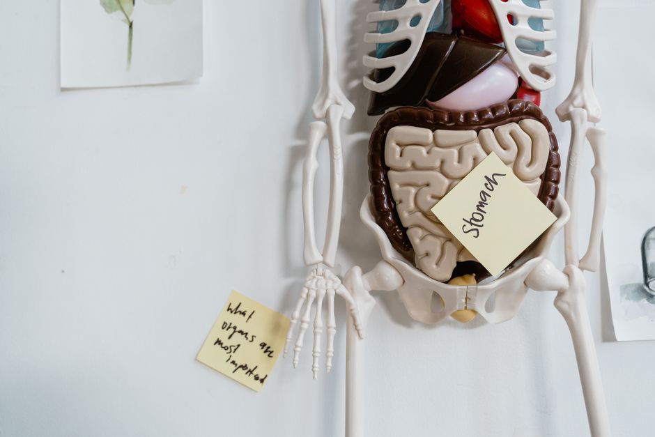
The digestive system is a long, muscular tube that runs from your mouth to your anus. When you look inside, you see a continuous pathway made of layers of tissue, blood vessels, and nerves. This tube, along with organs like the liver and pancreas, breaks down food into nutrients your body can use for energy, growth, and cell repair.
What Does The Digestive System Look Like Inside?
Picture a hollow tube about 30 feet long when fully grown. It is not a simple pipe. The walls are made of four distinct layers, each with a specific job. From the inside out, these layers are the mucosa, the submucosa, the muscularis, and the serosa.
The mucosa is the inner lining. It is moist, pink, and covered in tiny folds. In some areas, like the small intestine, these folds have finger-like projections called villi. These projections give the lining a velvety texture and massively increase the surface area for absorbing nutrients.
Beneath the mucosa lies the submucosa. This layer contains blood vessels, lymph vessels, and nerves. It supplies blood to the inner lining and carries absorbed nutrients away to the liver. The muscularis is the next layer, made of smooth muscle. It contracts in waves, a process called peristalsis, to push food forward. The outermost layer, the serosa, is a slippery membrane that lets the organs glide against each other inside your abdomen.
How Does Food Travel Through the Digestive Tract?
Digestion starts in the mouth. Your teeth tear and grind food into smaller pieces while saliva begins breaking down starches. The tongue shapes the food into a ball called a bolus and pushes it toward the throat.
The esophagus is a muscular tube about 10 inches long. It connects the throat to the stomach. A ring of muscle called the lower esophageal sphincter sits at the bottom of the esophagus. It opens to let food pass into the stomach and closes to prevent stomach acid from flowing back up.
Inside the stomach, the food mixes with acid and digestive enzymes. The stomach walls churn the mixture into a liquid called chyme. This organ looks like a deflated balloon when empty but can stretch to hold about a liter of food. The stomach lining is thick and covered in mucus to protect itself from the acid it produces.
After the stomach, chyme moves into the small intestine. This is where most digestion and absorption happen. It is about 20 feet long and coiled in the center of your abdomen. The first section, the duodenum, receives bile from the liver and enzymes from the pancreas. These fluids break down fats, proteins, and carbohydrates into molecules small enough to cross the intestinal wall.
The large intestine, or colon, is the final major section. It is about 5 feet long and wider than the small intestine. It frames the small intestine like a picture frame around a painting. The colon absorbs water and electrolytes from the remaining waste. It also houses trillions of bacteria that ferment undigested fiber and produce certain vitamins.
What Do the Accessory Organs Look Like and Do?
The liver, gallbladder, and pancreas are not part of the tube itself, but they are essential to digestion. The liver is the largest internal organ, weighing about 3 pounds. It sits on the right side of the abdomen, just below the diaphragm. It produces bile, a greenish-yellow fluid that breaks down fats.
The gallbladder is a small, pear-shaped sac tucked under the liver. It stores bile and releases it into the small intestine when you eat. The pancreas is a long, flat organ located behind the stomach. It produces digestive enzymes and releases them into the duodenum through a small duct.
These organs connect to the digestive tube through narrow ducts. The main pancreatic duct and the common bile duct join together before emptying into the duodenum at a point called the ampulla of Vater. This single opening delivers both bile and pancreatic enzymes to the food passing through.



How Long Does Food Stay in Each Part?
Transit time varies from person to person. It also depends on what you eat. In general, food spends about 2 to 4 hours in the stomach. Liquids pass through faster than solid foods. High-fat meals take longer to empty from the stomach than low-fat meals.
Movement through the small intestine takes about 3 to 5 hours. This slow passage gives nutrients time to be absorbed. The remaining material then enters the large intestine, where it can stay for 12 to 48 hours. Total transit time from mouth to toilet is typically between 24 and 72 hours.
Fiber affects transit time. Soluble fiber, found in oats and beans, forms a gel that slows digestion. Insoluble fiber, found in wheat bran and vegetables, adds bulk and speeds up movement through the colon. A diet with adequate fiber and fluids keeps the system moving at a healthy pace.
Why Does the Digestive System Look Different in Different People?
No two digestive systems look exactly alike. The length of the intestines varies from person to person. The position of the organs can shift slightly depending on body shape, posture, and whether you have had abdominal surgery.
Age changes the appearance of the digestive tract. The stomach lining thins with age in some people. The small intestine may absorb nutrients less efficiently. The colon can develop small pouches called diverticula, a condition called diverticulosis that becomes more common after age 60.
Previous surgery can change the anatomy. People who have had part of their stomach or intestine removed will have a shorter digestive tract. The remaining sections may adapt over time, but the overall structure will look different from someone who has never had surgery.
What Can You See With Medical Imaging?
Doctors use several tools to look inside the digestive system. An endoscope is a thin, flexible tube with a camera on the end. It is passed through the mouth to view the esophagus, stomach, and the first part of the small intestine. A colonoscope is similar but is inserted through the rectum to view the entire colon.
CT scans and MRI scans create detailed images of the digestive organs without entering the body. These scans show the shape, size, and position of the organs. They can reveal blockages, tumors, or areas of inflammation.
Barium studies involve drinking a chalky liquid that coats the inside of the digestive tract. The barium shows up white on X-rays, outlining the shape of the esophagus, stomach, and intestines. This technique highlights narrowing, ulcers, or abnormal pouches in the walls.
Frequently Asked Questions
How long is the entire digestive tract?
The digestive tract is about 30 feet long in a fully grown adult. Most of that length is the small intestine, which measures about 20 feet.
What color is the inside of the digestive system?
The inner lining is pink and moist, similar to the inside of your mouth. The color can vary slightly depending on blood flow and the specific organ.
Can you see your own digestive system?
You cannot see your digestive system without medical imaging or surgery. Doctors use endoscopy, CT scans, or MRI to view the inside of the tract.
Is the digestive system a single continuous tube?
Yes, it is one continuous tube from the mouth to the anus. The accessory organs like the liver and pancreas connect to this tube through small ducts.
