

Mycoplasma contamination is invisible to the naked eye and usually causes no obvious cloudiness in cell culture media. The first signs are often indirect: slowed cell growth, reduced transfection efficiency, or unexpected changes in cell behavior. Under a standard light microscope, contaminated cultures frequently look completely normal, which is why these infections can go unnoticed for weeks or months.
Why Mycoplasma Is So Hard to Spot
Mycoplasmas are the smallest self-replicating organisms known. They lack a cell wall, which makes them resistant to many common antibiotics used in cell culture, such as penicillin and streptomycin. They are also tiny — roughly 0.2 to 0.3 micrometers in diameter — which means they pass through standard 0.22-micrometer sterile filters used to keep media clean.
Because they are so small, they do not cause the turbidity that bacterial or fungal contamination typically produces. A flask of contaminated cells can look perfectly clear and healthy. The organisms attach to the outside of host cells or live in the spaces between them, drawing nutrients away without destroying the cells outright.
This silent behavior is what makes mycoplasma so dangerous in a laboratory. Cultures can carry the infection for months, contaminating experiments and producing unreliable data before anyone notices.
Early Warning Signs in Cell Culture
Most researchers first suspect mycoplasma when their cells start behaving oddly. The classic early indicators include:
- Slower cell growth and longer doubling times
- Reduced viability after thawing
- Decreased transfection efficiency
- Changes in protein expression patterns
- Unexpected shifts in cell morphology
- Spontaneous cell death or detachment
These symptoms are not specific to mycoplasma. Many other problems — nutrient depletion, passage number, or incubator issues — can produce similar changes. That is why routine testing is the only reliable way to confirm contamination.
Some strains cause more visible damage than others. Certain mycoplasma species deplete arginine from the media, which can slow DNA synthesis and cause cells to stop dividing. Others consume glucose or produce ammonia, gradually acidifying the media. Even then, the changes are subtle and easily mistaken for normal culture drift.
What Contamination Looks Like Under a Microscope
Under a standard light microscope, mycoplasma contamination is usually invisible. The organisms are far below the resolution limit of light microscopy. You cannot see individual mycoplasmas, and they do not form the colonies or clumps that bacteria and fungi create.
Some researchers report a fine granular appearance on the surface of infected cells, but this is not a reliable finding. It varies by cell line, mycoplasma species, and culture conditions. In most cases, the cells look entirely normal.
Electron microscopy can reveal mycoplasmas attached to cell membranes, but this is not practical for routine screening. The organisms appear as small, pleomorphic structures — often round, filamentous, or pear-shaped — clinging to the outer surface of host cells. This method is used for research confirmation, not everyday detection.
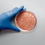




How Mycoplasma Contamination Is Actually Detected
Because visual inspection fails so consistently, laboratories rely on specific testing methods. The most common approaches include:
PCR testing is the fastest and most sensitive method. It detects mycoplasma DNA directly from culture supernatant or cells. Results are available within hours, and commercial kits can detect multiple species in a single test. PCR has become the standard screening tool in most research laboratories.
Hoechst or DAPI staining uses a fluorescent dye that binds to DNA. When viewed under a fluorescence microscope, contaminated cultures show small, punctate fluorescent spots outside the cell nuclei. These spots represent mycoplasma organisms. This method requires experience to interpret correctly, and false positives can occur if the stain is not handled properly.
Culture-based detection involves inoculating broth and agar media designed specifically for mycoplasma growth. This is the most sensitive method but takes weeks to produce results. It is used mainly for validating cell banks or confirming contamination in critical situations.
ELISA and other antibody-based tests detect mycoplasma antigens or antibodies in culture media. These are less sensitive than PCR but can be useful for screening large numbers of samples.
Most laboratories test new cell lines upon receipt, after thawing frozen stocks, and on a regular schedule — often monthly — for continuous cultures. Testing is also recommended whenever cells behave unexpectedly.
Common Sources of Mycoplasma in the Lab
Mycoplasma contamination almost always enters the laboratory through contaminated cell lines. The organisms spread easily between cultures through shared media, pipettes, and hoods. A single contaminated flask can infect an entire incubator within weeks.
Primary sources include:
- Cell lines received from other laboratories
- Frozen cell stocks that were contaminated before freezing
- Bovine serum used in media formulations
- Laboratory personnel (some mycoplasma species colonize humans)
- Contaminated reagents and trypsin solutions
Bovine serum is a well-documented source. Mycoplasmas can survive the filtration process used to sterilize serum, and some species naturally colonize cattle. Most commercial serum suppliers now screen for mycoplasma, but the risk is not zero, especially with serum from smaller suppliers.
Why Mycoplasma Contamination Ruins Experiments
Mycoplasma contamination does more than slow cell growth. It actively distorts experimental results in ways that can invalidate entire studies.
Infected cells show altered gene expression patterns. Mycoplasmas consume amino acids and nucleotides from the media, changing the metabolic state of the host cells. This can upregulate or downregulate specific genes, producing false positives or negatives in experiments measuring gene expression.
Immune responses are also affected. Mycoplasma components can activate Toll-like receptors and other inflammatory pathways in immune cells. This means experiments studying cytokine production, immune cell activation, or inflammatory signaling can produce completely misleading results.
Viral replication studies are especially vulnerable. Mycoplasma infection can interfere with viral entry, replication, and cytopathic effects. Some studies have shown that contaminated cells produce dramatically different viral titers compared to clean cells.
The most troubling aspect is that these effects are often subtle. An experiment may show a small but statistically significant difference between treatment groups, and the researcher may interpret this as a genuine biological effect when it is actually an artifact of mycoplasma infection.
Can Mycoplasma Contamination Be Treated?
Treatment of contaminated cultures is possible but not always successful. Several commercial antibiotic formulations are available, such as those containing fluoroquinolones or tetracycline derivatives. These drugs can eliminate mycoplasma from some cell lines.
The problem is that treatment is not always reliable. Some mycoplasma strains develop resistance. Some cell lines do not tolerate the treatment well and lose viability. And even when treatment appears successful, low-level contamination can persist and re-emerge later.
Most laboratories choose to discard contaminated cultures rather than treat them. This is the safer approach because it eliminates the risk of a persistent, low-grade infection that could compromise future experiments. If a cell line is irreplaceable — for example, a primary culture or a genetically modified line — treatment may be attempted, but the culture should be tested repeatedly afterward to confirm clearance.
Prevention is far more effective than treatment. Quarantining new cell lines, using mycoplasma-free reagents, practicing good aseptic technique, and testing regularly are the best defenses.
Frequently Asked Questions
Can you see mycoplasma contamination with a standard microscope?
No. Mycoplasmas are too small to be seen with a light microscope, and contaminated cultures usually look normal. Detection requires specific testing methods like PCR or fluorescent staining.
What are the first signs of mycoplasma contamination?
Slowed cell growth, reduced transfection efficiency, and unexpected changes in cell behavior are common early signs. These symptoms are not specific, so testing is needed to confirm the diagnosis.
How quickly does mycoplasma contamination spread between cultures?
Mycoplasma spreads rapidly through shared media, pipettes, and hoods. A single contaminated culture can infect an entire incubator within weeks if proper precautions are not followed.
Is it safe to treat mycoplasma-contaminated cells with antibiotics?
Treatment is possible but not always successful, and resistant strains can persist. Most laboratories discard contaminated cultures rather than risk ongoing experimental artifacts.
