
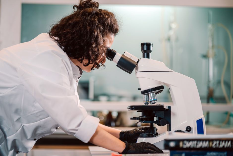
The microscope changed how we see the world in the most literal sense. Before it, humans knew that diseases existed, but had no way to see what caused them. The invention of the microscope made it possible to see a hidden universe of living things, including the cells that make up our bodies and the microbes that cause illness. This single tool opened the doors to modern biology, medicine, and our understanding of life itself.
What Did The Invention Of The Microscope Make Possible?
The invention of the microscope made it possible to observe objects and organisms that are invisible to the naked eye. This includes individual cells, bacteria, viruses, and the internal structures of plants and animals. Before the microscope, scientists could only study what they could see directly. After it, an entirely new biological world became available for study.
This was not just a matter of seeing smaller things. It changed the questions scientists could ask. Instead of guessing why food spoiled or why wounds became infected, researchers could actually look for the cause. The microscope turned biology from a descriptive science into an investigative one.
How Did the First Microscopes Work?
The earliest microscopes were simple devices with a single lens. Antonie van Leeuwenhoek, a Dutch draper, built some of the most powerful of these in the 1670s. His tiny glass spheres could magnify objects up to 270 times. With them, he became the first person to describe bacteria, red blood cells, and sperm cells.
Compound microscopes, which use multiple lenses, appeared around the same time. Robert Hooke used one to publish Micrographia in 1665, a book of detailed drawings of fleas, lice, and plant tissue. It was Hooke who first used the word “cell” to describe the tiny compartments he saw in a slice of cork. He chose the name because they reminded him of the small rooms monks lived in.
What Did the Microscope Reveal About Cells?
The microscope revealed that all living things are made of cells. This seems like basic knowledge now, but it took nearly 200 years of observation to establish. In 1839, two German scientists, Matthias Schleiden and Theodor Schwann, proposed the cell theory. It states that all plants and animals are composed of cells, and that the cell is the basic unit of life.
Later work added another key idea: all cells come from existing cells. This was proposed by Rudolf Virchow in 1855. The cell theory remains one of the most important foundations of biology. Every treatment for cancer, every vaccine, and every genetic test relies on the knowledge that life operates at the cellular level.
Modern microscopes have taken this much further. Electron microscopes can magnify objects up to millions of times, revealing the inner workings of cells in stunning detail. We can now see mitochondria, ribosomes, and even individual molecules. But every one of these advances traces back to the basic question first answered in the 1600s: what is actually there?





How Did the Microscope Change Medicine?
The microscope did more than reveal cells. It revealed the cause of infectious disease. Before the 1800s, many doctors believed that disease arose spontaneously from bad air or imbalance in the body. The microscope allowed researchers to see bacteria in infected tissue, water, and food.
Louis Pasteur used microscopes to show that fermentation was caused by living yeast cells. He also demonstrated that bacteria could spoil milk and wine, leading to the process of pasteurization. Robert Koch, working in the 1880s, used microscopes to identify the specific bacteria that cause tuberculosis, cholera, and anthrax. His work established the germ theory of disease, which is the foundation of modern medicine.
This understanding changed public health forever. Once doctors could see the bacteria in contaminated water, they could take action. Sewage systems, water treatment, and hand washing all became standard practice because of what the microscope revealed. Countless lives have been saved by these simple measures.
The microscope also made surgery safer. In the 1860s, Joseph Lister used the germ theory to develop antiseptic surgery. He sprayed carbolic acid on wounds and surgical instruments to kill bacteria. This dramatically reduced post-surgical infections and made operations far less deadly.
What Did the Microscope Reveal About Disease Diagnosis?
Today, the microscope remains an essential diagnostic tool. When a doctor suspects an infection, a sample of blood, urine, or tissue is placed on a slide and examined. This is how many bacterial infections, fungal infections, and parasitic diseases are confirmed.
A Pap smear, which screens for cervical cancer, involves examining cells from the cervix under a microscope. Pathologists look for abnormal changes that could indicate cancer or pre-cancerous conditions. Blood smears can reveal anemia, leukemia, or malaria. Biopsy samples from suspicious lumps are always examined microscopically to determine if they are cancerous.
Without the microscope, most of these diagnoses would be impossible. Doctors would be working blind, treating symptoms without knowing the underlying cause. The microscope provides the visual evidence needed to make informed treatment decisions.
What Did the Microscope Reveal About Genetics and DNA?
The microscope made it possible to see chromosomes, the structures that carry genetic information. In the late 1800s, scientists observed these thread-like structures inside dividing cells. They noticed that they appeared in pairs and were passed from parent cells to daughter cells.
This observation laid the groundwork for understanding heredity. In the early 1900s, Thomas Hunt Morgan used microscopes to study fruit flies and linked specific traits to specific chromosomes. This confirmed that genes are located on chromosomes, and that they control inherited characteristics.
Later, the microscope allowed scientists to see DNA itself. In 1953, James Watson and Francis Crick proposed the double helix structure of DNA, but it was Rosalind Franklin’s X-ray diffraction images that provided the evidence. These images, taken with a specialized microscope-like instrument, revealed the regular repeating pattern of the DNA molecule.
Today, fluorescent microscopes allow scientists to watch individual genes being expressed in living cells. This has transformed our understanding of genetic diseases, cancer, and development. The microscope turned genetics from a theoretical idea into a visible, testable science.
How Has the Microscope Advanced Modern Science?
Modern microscopes are far more powerful than anything Leeuwenhoek could have imagined. Electron microscopes use beams of electrons instead of light, allowing magnification of up to 10 million times. They can reveal the structure of viruses, the arrangement of atoms in a crystal, and the fine details of cell membranes.
Fluorescence microscopy uses dyes that glow under specific wavelengths of light. This allows scientists to label specific proteins or structures within a cell and watch them move in real time. It has been essential for understanding how cells divide, how neurons communicate, and how the immune system responds to infection.
In recent years, super-resolution microscopy has broken the traditional limits of light microscopy. These techniques allow scientists to see structures smaller than the wavelength of light, such as individual proteins and the tiny gaps between neurons. This has opened up entirely new areas of research in neuroscience and molecular biology.
What Could Not Have Been Discovered Without the Microscope?
Many of the most important discoveries in biology and medicine would have been impossible without the microscope. Consider this list:
- The existence of bacteria and viruses
- The cell theory and the understanding that all life is cellular
- The germ theory of disease and the development of vaccines
- The discovery of chromosomes and the basis of heredity
- The structure of DNA and the field of molecular biology
- The diagnosis of most cancers and blood disorders
- The development of antibiotics and their targeted use
None of these could have been achieved by observation with the naked eye alone. The microscope was not just a helpful tool. It was the essential instrument that made these fields of study possible.
Frequently Asked Questions
Who invented the first microscope?
Historians credit Zacharias Janssen, a Dutch spectacle maker, with building the first compound microscope around 1590, though his device was crude. Antonie van Leeuwenhoek later built powerful single-lens microscopes in the 1670s and was the first to observe bacteria and other microorganisms.
What was the first thing seen under a microscope?
Robert Hooke was the first to publish detailed observations of a sliced piece of cork, which he described as having small compartments he called “cells” in 1665. Antonie van Leeuwenhoek later became the first to observe living microorganisms in water, which he called “animalcules.”
Can modern microscopes see viruses?
Yes, electron microscopes can image viruses, which are typically too small to be seen with standard light microscopes. Fluorescence microscopy can also be used to detect viruses by labeling them with glowing markers.
Why is the microscope important in medicine today?
The microscope remains essential for diagnosing infections, cancers, and blood disorders by examining tissue and fluid samples. It is also used in research to develop new drugs, vaccines, and treatments by observing how cells respond to them.
