
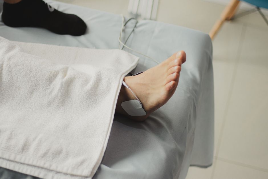
Baxter’s nerve entrapment happens when the first branch of the lateral plantar nerve gets compressed as it passes beneath the heel. The most common cause is chronic plantar fasciitis, where thickening and inflammation of the plantar fascia press against the nerve. Other causes include heel fat pad atrophy, bone spurs, and biomechanical overload from flat feet or high arches.
What Causes Baxters Nerve Entrapment?
Baxter’s nerve entrapment is a compression neuropathy. The nerve itself is not damaged by disease — it is being squeezed by surrounding tissue.
The first branch of the lateral plantar nerve (FBLPN) branches off the tibial nerve near the ankle and travels deep into the heel. Along its path, it passes between the abductor hallucis muscle and the medial calcaneal tuberosity — a narrow corridor where the nerve has very little room to move. Anything that narrows that corridor or thickens the surrounding tissue can compress the nerve.
This is why Baxter’s nerve entrapment rarely occurs in isolation. It is almost always secondary to something else happening in the foot.
What Is the Connection Between Plantar Fasciitis and Baxter’s Nerve Entrapment?
Chronic plantar fasciitis is the most common cause of Baxter’s nerve entrapment. Research consistently shows that a large share of people diagnosed with chronic heel pain have both conditions at the same time.
The mechanism is mechanical. When the plantar fascia becomes chronically inflamed, it thickens. That thickened fascia — along with the swelling and scar tissue that builds up over months — narrows the space where the nerve runs. The nerve gets compressed between the thickened fascia and the muscle above it.
This is why some people with plantar fasciitis do not improve with standard treatment. Stretching, orthotics, and anti-inflammatory medication address the fascia. They do not decompress the nerve. If the nerve is the dominant pain source, those treatments will not fully resolve the problem.
The overlap is significant enough that some clinicians evaluate for nerve involvement in any case of heel pain lasting more than a few months. That is common clinical practice, though it is not a formal evidence-based recommendation.
What Role Does Heel Fat Pad Atrophy Play?
The heel fat pad is a specialized cushion of fatty tissue that absorbs impact with every step. It is not ordinary fat. It has a honeycomb structure of fibrous bands that hold the fat in place and allow it to deform under load then spring back.
With age, or after repeated corticosteroid injections into the heel, that structure can break down. The pad gets thinner. The heel loses its natural shock absorber.
When the fat pad atrophies, the mechanical forces that would normally be absorbed by the cushion get transferred to the structures underneath — including the nerve. The nerve is not designed to absorb impact. Repeated compression from walking and standing can irritate it over time.
This is one reason why corticosteroid injections into the heel are used cautiously. They reduce inflammation in the short term, but repeated injections are associated with fat pad atrophy and, in some cases, plantar fascia rupture. The evidence on how often this occurs is mixed, but the risk is well recognized in clinical practice.





Can Bone Spurs Cause Baxter’s Nerve Entrapment?
Bone spurs — calcifications at the heel where the plantar fascia attaches — are commonly seen on X-rays of people with heel pain. But their role in causing nerve compression is more limited than many people assume.
Many people with heel spurs have no pain at all. And many people with Baxter’s nerve entrapment have no spur. The presence of a spur on imaging does not confirm it is the pain source.
That said, a spur located near the nerve’s path can contribute to compression, especially when combined with other factors like fascia thickening or fat pad thinning. It is one piece of the puzzle, not the whole picture.
This is a point where imaging can mislead. A finding on an X-ray or MRI does not automatically mean that finding is causing the symptoms.
How Do Foot Mechanics Contribute to Nerve Compression?
Foot structure affects how load is distributed through the heel. Two opposite problems can both lead to nerve compression.
- Flat feet (overpronation): The arch collapses with each step, which stretches the plantar fascia and alters the alignment of the muscles around the heel. This can narrow the space around the nerve.
- High arches (cavus feet): The arch is rigid and does not absorb shock well. Impact forces concentrate on the heel, increasing pressure on the nerve.
- Excessive pronation during walking or running: Even without flat feet, repeated inward rolling of the foot can stretch and irritate the nerve over time.
Biomechanical factors rarely cause Baxter’s nerve entrapment on their own. They create the conditions — increased pressure, altered alignment, repetitive strain — that make compression more likely when combined with other causes.
Which Activities and Occupations Increase the Risk?
Anything that puts repeated load on the heel increases the risk of nerve compression.
Running — especially on hard surfaces or in worn-out shoes — is a common trigger. So is any job that requires prolonged standing or walking on hard floors. Nurses, factory workers, restaurant staff, and retail employees are among those who spend hours on their feet daily.
Being overweight increases the load through the heel with every step. The relationship between body weight and heel pain is well established, though the exact contribution to nerve entrapment specifically is less clear.
Sudden increases in activity — going from sedentary to running five miles, or starting a job that requires standing after years at a desk — can also provoke symptoms. The nerve tolerates gradual load increases better than sudden ones.
What Other Conditions Can Cause Similar Heel Pain?
Baxter’s nerve entrapment is often confused with other causes of heel pain because the symptoms overlap. Getting the diagnosis right matters because the treatments differ.
| Condition | Key Distinguishing Feature |
|---|---|
| Plantar fasciitis | Pain worst with first steps in the morning, improves with movement |
| Baxter’s nerve entrapment | Pain that burns, tingles, or radiates; may worsen with activity rather than improve |
| Tarsal tunnel syndrome | Pain or numbness along the inside of the ankle, not just the heel |
| Stress fracture of the calcaneus | Pain that worsens with weight-bearing and does not improve with rest |
| Fat pad atrophy | Pain directly under the heel bone, worse on hard surfaces |
In practice, many people have more than one of these at the same time. That is part of why heel pain can be so stubborn.
Why Is Baxter’s Nerve Entrapment Often Missed?
It is frequently overlooked because plantar fasciitis is so much more common. When someone walks into a clinic with heel pain, the default assumption is plantar fasciitis. If the initial treatment does not work, the next step is often more aggressive plantar fasciitis treatment rather than reconsidering the diagnosis.
Baxter’s nerve entrapment also does not have a single definitive test. Diagnosis relies on clinical examination — the location of tenderness, the quality of pain, and response to specific maneuvers — combined with imaging when needed. Nerve conduction studies and MRI can support the diagnosis but are not always conclusive.
The practical takeaway: if heel pain has not improved after several months of appropriate treatment for plantar fasciitis, it is reasonable to ask whether nerve involvement is part of the picture.
Frequently Asked Questions
What is the most common cause of Baxter’s nerve entrapment?
Chronic plantar fasciitis is the most common cause, because thickening of the plantar fascia compresses the nerve where it passes beneath the heel. Heel fat pad atrophy and biomechanical overload are also significant contributors.
How is Baxter’s nerve entrapment different from plantar fasciitis?
Plantar fasciitis typically causes pain that is worst with the first steps in the morning and improves with movement. Baxter’s nerve entrapment tends to cause burning, tingling, or radiating pain that may worsen with activity rather than improve.
Can Baxter’s nerve entrapment go away on its own?
Mild cases may improve with rest, activity modification, and footwear changes, but the evidence on spontaneous recovery is limited. Persistent or worsening symptoms should be evaluated by a clinician.
Does Baxter’s nerve entrapment show up on an MRI?
MRI can show signs of nerve inflammation or surrounding tissue changes, but it does not always confirm the diagnosis. Clinical examination remains the primary diagnostic tool.
