
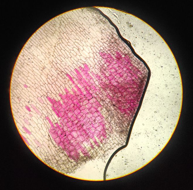
Collagen fibers are the most abundant protein structures in the human body, and in histology they appear as wavy, pink-staining bundles when tissue is treated with standard dyes. They give skin, tendons, bones, and organs their tensile strength. When a pathologist looks at a tissue sample under a microscope, collagen fibers are often the first structural element they identify to understand what kind of tissue they are examining.
What Are Collagen Fibers In Histology?
In histology, collagen fibers are extracellular structures made primarily of type I collagen protein. They form when triple-helix collagen molecules assemble into fibrils, and those fibrils bundle together into fibers visible under a light microscope.
The word “collagen” comes from the Greek words for “glue producer.” That name reflects a basic truth: collagen holds tissues together. It is the scaffolding of the body.
On a stained slide, collagen fibers typically appear as:
- Wavy, undulating bands
- Pink or red with hematoxylin and eosin (H&E) staining
- Blue or green with trichrome stains
- Variable in thickness depending on the tissue and the type of collagen present
Collagen is not a single substance. At least 28 types of collagen have been identified in humans. Type I is by far the most common and the one most relevant to histology. It dominates in skin, bone, tendon, ligaments, and the fibrous capsules around organs.
Type II collagen is found mainly in cartilage. Type III collagen is common in blood vessels, skin, and early wound healing tissue. Type IV forms the meshwork of basement membranes. Each type has a distinct distribution and role, and histologists use that distribution to identify tissues and detect disease.
How Do Collagen Fibers Appear Under a Microscope?
Collagen fibers are visible under a light microscope after standard tissue staining. They do not absorb most dyes the way cells do, so their appearance depends heavily on the stain used.
With H&E staining — the most common stain in pathology — collagen fibers appear pink (eosinophilic). They look like broad, wavy ribbons running through the extracellular space. Cell nuclei stain blue or purple, which creates contrast. This contrast is what lets a pathologist distinguish collagen from muscle, fat, or connective tissue cells.
With trichrome stains, the picture changes. These stains are designed to differentiate collagen from muscle and cytoplasm. Collagen typically stains blue or green, while muscle and cytoplasm stain red or pink. Trichrome staining is often used when the amount or distribution of collagen matters — for example, in liver biopsies or kidney biopsies.
Under polarized light, collagen fibers appear birefringent. That means they split light into two rays with different refractive indices. This property reflects the highly ordered arrangement of collagen molecules within the fiber. It is not just a lab curiosity — polarized light microscopy is sometimes used to help identify collagen in ambiguous samples.
Under an electron microscope, the hierarchical structure becomes clear. Individual collagen molecules assemble into fibrils with a characteristic banding pattern. Those fibrils group into fibers. The fibers group into larger bundles. This organization is what gives collagen its strength.
What Types of Collagen Exist and Where Are They Found?
Collagen types differ in their amino acid sequence, their assembly, and their location in the body. The table below summarizes the most studied types.
| Collagen Type | Primary Location | Key Feature |
|---|---|---|
| Type I | Skin, bone, tendon, ligaments, dentin | Most abundant; forms thick, strong fibers |
| Type II | Cartilage (hyaline and elastic) | Forms fine fibrils; provides cartilage resilience |
| Type III | Blood vessels, skin, uterus, early wound tissue | Thinner fibers; often co-localizes with type I |
| Type IV | Basement membranes | Forms a sheet-like network, not fibrils |
| Type V | Cornea, bone, placenta | Regulates fibril diameter |
This list is not exhaustive. Other types play important roles in specific tissues. But these five are the ones most commonly discussed in histology and pathology.
One clarification worth making: not all collagen forms fibers. Type IV collagen, for example, assembles into a mesh-like sheet rather than fibrils. When histologists talk about “collagen fibers,” they are usually referring to the fibril-forming types — mainly I, II, III, V, and XI.





How Is Collagen Built in the Body?
Collagen synthesis is a multi-step process that starts inside cells and finishes outside them. Understanding this process helps explain why certain diseases affect collagen and why nutritional deficiencies can cause problems.
Here is the sequence in simplified form:
- Collagen genes are transcribed and translated into polypeptide chains called preprocollagen.
- Inside the cell, enzymes modify the chains by adding hydroxyl groups to certain amino acids. Vitamin C is required for this step.
- Three chains twist together into a triple helix, forming procollagen.
- Procollagen is secreted from the cell.
- Outside the cell, enzymes trim the ends of the molecule, converting it to tropocollagen.
- Tropocollagen molecules self-assemble into fibrils.
- Cross-linking between molecules stabilizes the fibrils and gives collagen its tensile strength.
The vitamin C connection is well established. Without adequate vitamin C, the hydroxylation step fails, and collagen cannot form properly. This is the mechanism behind scurvy — a condition characterized by poor wound healing, bleeding gums, and fragile blood vessels. Scurvy is rare in the United States but still occurs in people with severe malnutrition or restricted diets.
Cross-linking is another critical step. It involves enzymes that form strong bonds between collagen molecules. As people age, cross-linking patterns change, which affects tissue stiffness and elasticity. This is one reason why arteries stiffen and skin loses some of its flexibility over time.
What Happens When Collagen Goes Wrong?
Collagen disorders fall into two broad categories: genetic conditions that affect collagen production or structure, and acquired conditions where collagen is damaged, overproduced, or deposited abnormally.
Genetic collagen disorders include several well-characterized conditions. Osteogenesis imperfecta is caused by mutations in type I collagen genes and leads to fragile bones. Ehlers-Danlos syndromes are a group of conditions affecting collagen and related connective tissue proteins, with symptoms that can include joint hypermobility and skin that bruises or stretches easily. Some forms of chondrodysplasia affect type II collagen and result in cartilage and bone abnormalities.
These conditions vary widely in severity. Some are mild and only noticed in adulthood. Others are evident at birth. Diagnosis typically involves clinical evaluation, family history, and sometimes genetic testing. No single treatment corrects the underlying collagen defect, and management focuses on preventing complications.
Acquired collagen changes are more common. Fibrosis is the excessive deposition of collagen in an organ, often after injury or chronic inflammation. It occurs in the liver (cirrhosis), lungs (pulmonary fibrosis), kidneys, heart, and other tissues. In fibrosis, collagen that normally provides structure accumulates beyond what is needed, replacing functional tissue and impairing organ function.
Scar formation after wound healing is another example. Collagen is laid down to close the wound, but the new tissue does not perfectly replicate the original. In some people, scars become raised and thickened — a condition called keloid scarring. The exact reasons why some people form keloids and others do not are not fully understood, though genetics appear to play a role.
How Do Histologists Identify Collagen in Tissue Samples?
Identifying collagen in a tissue sample requires more than seeing pink fibers. Experienced histologists and pathologists use a combination of staining patterns, tissue architecture, and sometimes specialized techniques.
The first step is usually H&E staining. Collagen’s pink, wavy appearance is often enough to recognize it in routine samples. But H&E does not always distinguish collagen from other eosinophilic structures like muscle or fibrin.
When more certainty is needed, special stains help:
- Masson’s trichrome stains collagen blue or green, making it easy to distinguish from muscle and cytoplasm.
- Van Gieson’s stain stains collagen red and muscle yellow.
- Reticulin stain highlights type III collagen fibers, which form delicate networks in organs like the liver and spleen.
- Immunohistochemistry uses antibodies that bind to specific collagen types, allowing precise identification.
Polarized light microscopy can also help. Because collagen fibers are birefringent, they appear bright against a dark background when viewed under polarized light. This technique is sometimes used to confirm collagen in tissue or to assess its organization.
In research settings, electron microscopy reveals the characteristic banding pattern of collagen fibrils. This level of detail is rarely needed for clinical diagnosis but is valuable for understanding collagen structure and disease mechanisms.
Why Does Collagen Matter in Medicine?
Collagen matters in medicine because it is involved in so many normal and abnormal processes. When collagen is defective, too little, or too much, the consequences can be serious.
In surgery, collagen integrity affects how tissues heal. Surgeons know that tissues with poor collagen — whether from disease, malnutrition, or aging — may heal more slowly or with weaker scars. This is one reason why nutritional status is assessed before major operations.
In rheumatology and orthopedics, collagen is central to conditions like osteoarthritis, where cartilage (rich in type II collagen) breaks down over time. In dermatology, collagen changes are linked to skin aging and scarring. In cardiology, collagen in heart valves and arteries affects how those structures function.
Collagen is also used in medical products. Collagen-based dressings, sutures, and implants are common. Some are derived from animal sources, while others are synthetic. These products are regulated as medical devices, and their safety and effectiveness are evaluated by regulatory agencies.
What collagen supplements can and cannot do is a separate question. Oral collagen supplements are widely marketed for skin, joint, and hair health. The evidence here is mixed. Some studies suggest that certain collagen peptides may improve skin elasticity or joint comfort, but results vary, and many studies are small or industry-funded. No clinical evidence currently confirms that oral collagen supplements rebuild collagen fibers in the skin or joints in a way that produces meaningful clinical benefit for everyone.
Frequently Asked Questions
What do collagen fibers look like in histology?
Collagen fibers appear as wavy, pink-staining bundles under H&E staining and blue or green with trichrome stains. Their appearance varies with the stain and the type of collagen present.
What is the difference between collagen fibers and collagen fibrils?
Collagen fibrils are smaller structures that bundle together to form fibers. Fibrils are visible under electron microscopy, while fibers are visible under light microscopy.
Which collagen type is most common in the human body?
Type I collagen is the most abundant, found in skin, bone, tendon, ligaments, and many other tissues. It accounts for roughly 90% of the body’s total collagen, though exact figures vary by source and method of measurement.
Can collagen fibers regenerate after injury?
Collagen fibers can be repaired and replaced after injury, but the new tissue often differs from the original in structure and strength. Complete regeneration without scar formation is limited in most adult tissues.
