
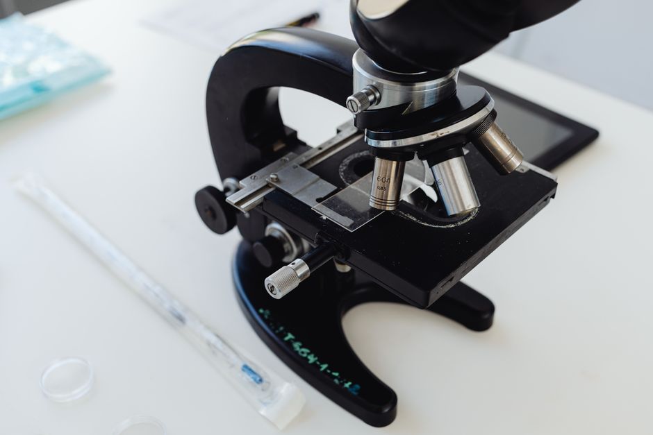
Reading a urinalysis dipstick and microscopy result means looking at three things: color and clarity, chemical markers on the dipstick, and cells or crystals under a microscope. The dipstick checks for things like protein, glucose, blood, and infection markers in seconds. The microscopy looks for red blood cells, white blood cells, bacteria, and casts that the dipstick might miss. Together they give a full picture of what is in the urine and what it might mean for your health.
What Do the Dipstick Test Strip Colors Actually Mean?
The dipstick is a plastic strip with small colored squares. Each square tests for a different substance. You dip the strip into fresh urine, wait the time listed on the bottle, and compare the colors to a chart on the label.
Here is what the common squares check for:
- Leukocytes — turns purple if white blood cells are present. This can mean a urinary tract infection.
- Nitrite — turns pink if bacteria that turn nitrate into nitrite are in the urine. This is also a sign of infection.
- Protein — changes color if protein is leaking into urine. Small amounts can be normal. Higher levels may point to kidney issues.
- Glucose — turns positive when blood sugar is high enough to spill into urine. This usually means diabetes or prediabetes.
- Ketones — shows up when the body burns fat for energy instead of sugar. Common in fasting, very low carb diets, or uncontrolled diabetes.
- Blood — detects red blood cells or hemoglobin. Can be from infection, kidney stones, or menstruation.
- pH — measures how acidic or alkaline the urine is. Normal range is roughly 4.5 to 8. Diet and medications affect this.
- Specific gravity — tells how concentrated the urine is. High means dehydrated. Low means overhydrated or possible kidney problems.
- Urobilinogen — a breakdown product of bilirubin. High levels can point to liver issues.
- Bilirubin — should be zero. Any positive result needs follow-up for possible liver or bile duct problems.
The key is reading the timing correctly. If you check too early or too late the color may be wrong. Always follow the exact seconds listed on the strip bottle.
How Do You Interpret Microscopy Results?
After the dipstick, a lab technician spins the urine in a centrifuge. This concentrates the solid material at the bottom. They put a drop of that sediment on a slide and look at it under a microscope.
Microscopy is more detailed than the dipstick. It can find things the chemical strip misses. Here is what they are looking for:
- Red blood cells — more than 2-3 per high power field is abnormal. Can come from infection, stones, trauma, or kidney disease.
- White blood cells — more than 5 per high power field suggests inflammation or infection.
- Bacteria — if present with white blood cells it strongly suggests a UTI. Without white cells it could be contamination from collection.
- Epithelial cells — small numbers are normal. Large numbers mean a poor sample or possible vaginal contamination.
- Casts — these are tube-shaped clumps of protein or cells. They form in the kidney tubules. Red cell casts mean kidney bleeding. White cell casts mean kidney inflammation. Hyaline casts can be normal with dehydration.
- Crystals — common types include calcium oxalate, uric acid, and struvite. Some are normal. Large amounts or specific shapes may point to kidney stones or metabolic disorders.
Normal results show few cells and no casts or bacteria. Abnormal results need correlation with symptoms and other lab work.
How To Read A Ua Dipstick And Microscopy Results Together
Reading each part alone can be misleading. The dipstick and microscopy complement each other. You need to cross-check them.
For example, a dipstick positive for blood but no red cells on microscopy can mean hemoglobin or myoglobin is present. That happens with muscle breakdown or certain blood disorders. A dipstick negative for leukocytes but microscopy showing white cells could mean the infection is mild or the strip expired.
Here is a comparison of what the two methods detect:
| Finding | Dipstick | Microscopy |
|---|---|---|
| Red blood cells | Detects blood chemically | Counts actual cells |
| White blood cells | Detects leukocyte enzyme | Counts actual cells |
| Bacteria | Nitrite test only catches some | Sees bacteria directly |
| Protein | Good screening tool | Cannot quantify |
| Casts | Cannot detect | Only way to see them |
| Crystals | Cannot detect | Only way to see them |
If the dipstick and microscopy disagree, the microscopy is usually more reliable for cells and casts. The dipstick is better for chemicals like glucose and protein.




What Can Throw Off Urinalysis Results?
Many things can make a urinalysis inaccurate. Knowing them helps you avoid false alarms.
Contamination is the most common problem. Bacteria or cells from the skin or vagina can get into the sample. This is why a clean catch midstream sample matters. The first bit of urine flushes out contaminants. Collect the middle part.
Timing matters. Urine that sits at room temperature for more than an hour changes. Bacteria multiply. Cells break down. Crystals form or dissolve. The lab should process the sample within 60 minutes or refrigerate it.
Medications and supplements can change colors on the dipstick. Vitamin C can cause false negatives for blood and glucose. Certain antibiotics can interfere with leukocyte and nitrite tests. Always tell your doctor what you take.
Menstruation can cause blood on the dipstick and red cells on microscopy. This is normal during a period. If you are menstruating, tell the lab or wait until it ends.
Dehydration concentrates the urine. This can make protein and specific gravity look higher than they really are. Drinking too much water dilutes the urine and can hide problems.
The American Association for Clinical Chemistry notes that dipstick results are screening tests only. Abnormal results need confirmation with more specific tests.
What Does Research on Urinalysis Accuracy Show?
Research published in the Clinical Journal of the American Society of Nephrology found that dipstick protein testing has about 80-90% sensitivity for detecting significant proteinuria. That means it misses 10-20% of cases. Microscopy catches some of those missed cases when casts are present.
A study in Journal of Clinical Microbiology looked at dipstick nitrite and leukocyte tests for UTIs. The combination had a sensitivity of about 75% compared to culture. That means one in four infections could be missed if only the dipstick is used. Adding microscopy improved detection.
The CDC recommends urinalysis as a first step but not a final diagnosis for kidney disease or infection. Abnormal results always need follow-up testing like urine culture, blood work, or imaging.
Some people claim that cloudy or smelly urine always means infection. That is not true. Cloudiness can come from crystals or vaginal discharge. Smell can come from food like asparagus. The dipstick and microscopy are needed to tell the difference.
What Do Normal vs Abnormal Results Look Like?
Here is a quick reference for what typical results look like:
Normal urinalysis: Clear or pale yellow. No protein, glucose, ketones, blood, nitrite, or leukocytes on dipstick. pH around 6. Specific gravity 1.005 to 1.030. Microscopy shows fewer than 2 red cells, fewer than 5 white cells, occasional epithelial cells, and no casts or bacteria.
Possible UTI: Cloudy urine. Dipstick positive for leukocytes and nitrite. Microscopy shows many white blood cells and bacteria. Red cells may be present.
Possible kidney disease: Dipstick positive for protein and possibly blood. Microscopy shows red cell casts or fatty casts. Protein may be high enough to cause foamy urine.
Possible diabetes: Dipstick positive for glucose. Ketones may also be present if blood sugar is very high. Specific gravity may be high due to glucose pulling water out.
Possible kidney stones: Dipstick positive for blood. Microscopy shows red blood cells and crystals. Crystals may be calcium oxalate or uric acid depending on stone type.
These patterns are not guarantees. They are starting points for further investigation.
Common Misconceptions About Urinalysis
Many people think a dipstick alone is enough to diagnose a UTI. It is not. The nitrite test misses bacteria that do not convert nitrate, like enterococcus. The leukocyte test can be negative early in infection. Culture is the gold standard.
Some believe that trace protein on dipstick is always serious. It is often benign and temporary from exercise, fever, or stress. Persistent protein needs follow-up. A single trace reading does not mean kidney disease.
Another myth is that dark urine means you are dying. Dark urine can come from dehydration, beets, rhubarb, or certain medications. It can also mean blood or bilirubin. You need the dipstick and microscopy to know which.
There is a viral claim that bubbles in urine mean kidney failure. Foamy urine can mean protein, but it can also just mean the urine hit the water fast. The dipstick protein test tells you if it is real protein or just air bubbles.
Frequently Asked Questions
How long does a urinalysis dipstick take to read?
Most dipsticks need 60 to 120 seconds for accurate color changes. Read the specific timing on the bottle you are using.
Can you read a urinalysis dipstick at home?
Yes, home dipstick kits are available. They work the same way as lab strips but are less precise. Always confirm abnormal results with a doctor.
What does trace blood on a dipstick mean?
Trace blood can be from a mild infection, kidney stone, exercise, or menstruation. It needs microscopy to check for actual red blood cells.
Do you need to fast before a urinalysis?
No fasting is required. Avoid large amounts of vitamin C and beets beforehand as they can change dipstick colors.
