
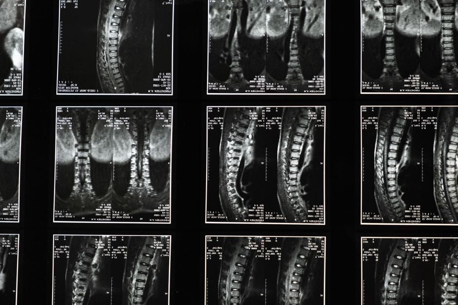
Cervical instability is a condition where the neck’s ligaments are too loose to hold the vertebrae in proper alignment. Diagnosis relies on a combination of your medical history, a physical exam, and specific imaging tests. Doctors typically start with standard X-rays and MRI, then may order flexion-extension X-rays to see how the bones move when you bend your neck forward and backward.
What Is Cervical Instability?
The cervical spine consists of seven vertebrae stacked from the skull base to the upper back. Ligaments and muscles keep these bones stable while allowing normal movement. Cervical instability means these supporting structures have become too lax. The vertebrae can shift more than they should, sometimes irritating nerves or the spinal cord itself.
This excessive movement can happen after trauma such as whiplash, from connective tissue disorders, or after surgery. Some people are born with naturally looser ligaments. The condition is not always easy to identify because symptoms overlap with many other neck problems.
What Are the Common Symptoms of Cervical Instability?
Symptoms vary widely from person to person. Many report neck pain, headaches that start at the base of the skull, and a sensation that the head feels heavy or hard to hold up. You might hear or feel clicking and grinding when turning your head.
Some people experience dizziness, blurred vision, or ringing in the ears. Others have tingling or numbness in the arms and hands. When the spinal cord is irritated, symptoms can include problems with balance, bladder control, or weakness in the arms and legs. These symptoms are not unique to cervical instability, which is why diagnosis takes careful evaluation.
Who Is at Risk for Cervical Instability?
Anyone can develop cervical instability, but certain groups are at higher risk. People who have been in car accidents, especially those with whiplash injuries, often damage the neck ligaments. Athletes in contact sports like football or rugby also face increased risk.
Connective tissue disorders such as Ehlers-Danlos syndrome make ligaments inherently weaker. People with rheumatoid arthritis can develop instability from inflammation that loosens ligaments. Prior neck surgery, especially multiple fusions, can shift stress to nearby segments and cause instability over time.





How Is Cervical Instability Diagnosed: Tests and Imaging?
Diagnosis starts with a detailed history. Your doctor will ask about past injuries, surgeries, and specific symptoms. Next comes a physical exam where the doctor checks your neck range of motion, strength, reflexes, and signs of nerve irritation.
Imaging is the next step. Standard X-rays show bone alignment and can reveal abnormal spacing between vertebrae. MRI provides detailed images of soft tissues including ligaments, discs, and the spinal cord. It can show if ligaments are torn or stretched. CT scans give a clearer view of bone structure and are sometimes used when X-rays or MRI are not enough.
Flexion-extension X-rays are often the key test. These are two sets of X-rays taken with your neck bent fully forward and then fully backward. The radiologist measures how much the vertebrae slide relative to each other. More than 3.5 mm of movement between two vertebrae is considered abnormal in adults. Some specialists also use digital motion X-ray, which captures video of the neck moving in real time. This can show subtle instability that static X-rays miss.
In some cases, a CT myelogram is ordered. This involves injecting contrast dye into the spinal canal and then taking CT images. It can reveal if the spinal cord is being compressed during movement. Provocative discography is sometimes used but remains controversial because it is invasive and not always reliable.
What Does the Physical Exam Look For?
The physical exam is an essential part of the diagnosis. Your doctor will watch how you move your neck and note any painful arcs or limitations. They will press on the spine and the muscles beside it to find tender spots.
Neurological testing checks muscle strength in your arms and legs, reflexes, and sensation to light touch and pinprick. Tests like the Hoffman sign test for a reflex that can indicate spinal cord irritation. The doctor may also perform a Sharp-Purser test, where they gently push your forehead backward while stabilizing the second vertebra. If this reproduces your symptoms, it suggests instability at the upper neck level.
What Are the Limitations of Current Imaging Techniques?
No single test confirms cervical instability with complete certainty. Standard X-rays and MRI taken while you lie still do not show how the bones move. Flexion-extension X-rays help, but patient effort and pain can limit how much a person bends, leading to false normal results.
MRI can show ligament damage, but not all torn ligaments cause instability. Some people have abnormal MRI findings without symptoms. Conversely, functional instability can exist with a normal MRI. Many people have mild degrees of abnormal movement without problems. The diagnosis depends on linking the imaging findings to your specific symptoms.
Some newer imaging techniques, such as upright MRI or dynamic CT, allow scanning while the neck is loaded by gravity. These are not yet widely available, and evidence supporting their routine use is still emerging. The decision to use advanced imaging should be made with a specialist who understands the nuances.
When Should You See a Specialist?
If you have persistent neck pain, headaches, or neurological symptoms after an injury, consider seeing a spine specialist. A neurosurgeon or an orthopedic surgeon who focuses on the spine is best equipped to evaluate cervical instability. Physiatrists and pain management doctors also manage this condition.
Before seeing a specialist, keep a record of your symptoms. Note when they started, what triggers them, and what makes them better. Bring previous imaging reports and any surgery records. This information helps the doctor decide which tests are most appropriate for your case.
Frequently Asked Questions
What is the best test for cervical instability?
There is no single best test. Most doctors rely on flexion-extension X-rays to measure abnormal movement between vertebrae. MRI provides important information about ligament and nerve health, and a thorough history and physical exam are equally critical.
Can cervical instability be seen on MRI?
MRI can show torn or stretched ligaments, which can suggest instability, but it does not directly measure movement. A normal MRI does not rule out instability if symptoms and exam findings are strong. Dynamic imaging (flexion-extension) is usually needed to confirm the diagnosis.
How long does it take to diagnose cervical instability?
Diagnosis can take weeks to months because symptoms mimic many other conditions and because the right imaging must be ordered and interpreted. Getting a proper evaluation by a spine specialist often speeds up the process.
Is cervical instability curable?
Many people improve with physical therapy, activity modification, and bracing. When conservative treatments fail and instability is severe, surgery to fuse the unstable vertebrae can resolve symptoms. The term “cure” is rarely used because the underlying ligament weakness may persist, but treatment is often effective.
