
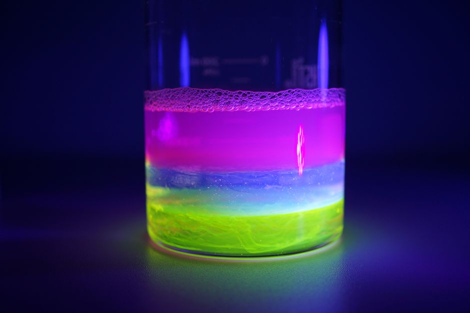
RNA is invisible under a standard microscope, yet researchers can watch it move inside living cells. They do it by attaching fluorescent dyes that light up when they bind RNA. The dye absorbs light at one wavelength and re-emits it at a longer one, so a microscope tuned to that emitted light sees the RNA as a bright glow against a dark background. That simple physical trick is the basis for much of what scientists know about where RNA is made, where it travels, and how much of it exists in a given cell.
Why Is RNA Invisible Without a Fluorescent Label?
RNA does not absorb or emit visible light in any useful way. It is a chain of nucleotides, and under ordinary light microscopy it is essentially transparent. The same is true of DNA and most proteins. To see any of them, you have to give them something that interacts with light.
Older methods relied on colored stains that bind nucleic acids. Those stains can show that RNA is present in a cell, but they cannot tell you which specific RNA it is, and they often cannot distinguish RNA from DNA. Fluorescent dyes solved both problems. A dye molecule absorbs photons at one wavelength, gets pushed into a higher energy state, then releases that energy as a photon at a longer wavelength. A microscope filters out the incoming light and collects only the emitted light. The result is a high-contrast image where the labeled target appears bright.
The key advantage is specificity. A dye attached to a probe that recognizes one RNA sequence will light up only that sequence. This is why fluorescence became the standard tool for studying RNA rather than a single generic stain.
How Fluorescent Dyes Make RNA Visible: The Basic Mechanism
Fluorescence is a two-step physical process. A fluorophore absorbs a photon and its electrons jump to an excited state. Within nanoseconds, some energy is lost as heat, and the electron falls back to a lower state, releasing a photon of lower energy and longer wavelength. That gap between absorbed and emitted light is called the Stokes shift, and it is what lets a microscope separate signal from background.
To make RNA visible, the dye must end up bound to RNA and nowhere else. There are three common ways this happens.
- Sequence-specific probes. A short piece of nucleic acid complementary to the target RNA carries a fluorophore. When it binds its target, the dye is now localized to that RNA. Unbound probes are washed away or designed to stay dark.
- Intercalating and groove-binding dyes. Some dyes slip between base pairs or sit in the minor groove of double-stranded RNA. They fluoresce strongly only when bound, so free dye in solution stays dim.
- Fluorogenic probes. These are designed to be dark until they contact their target. A common design keeps a quencher next to the fluorophore; when the probe binds RNA, the quencher moves away and the dye lights up. This reduces background dramatically.
The common thread is that the dye must be switched on by binding. A dye that glows everywhere tells you nothing about RNA location.
What Kinds of Fluorescent Dyes Are Used for RNA?
Different dyes suit different questions. Some are workhorses for imaging fixed cells, others are designed for live cells, and a few are built for single-molecule detection.
For fixed cells, dyes that bind nucleic acids broadly are common. They reveal total RNA distribution but not identity. For sequence-specific work, researchers attach a fluorophore to an antisense probe, a molecular beacon, or a similar targeting molecule. For live-cell imaging, the challenge is that many dyes are toxic or get pumped out of cells, so dyes with better retention and lower toxicity are preferred.
Green and red emitting dyes are the most widely used because standard microscopes and filter sets handle them well. Far-red dyes are useful for thick samples because longer wavelengths scatter less and penetrate tissue better. A practical point that is easy to miss: the choice of dye is often limited less by the RNA target and more by the microscope, the filter sets available, and whether the cells must stay alive.




How Do Researchers Attach Dyes to Specific RNA Sequences?
Attaching a dye to a specific RNA is the hard part. The dye itself does not know which RNA to find. That job belongs to a targeting molecule.
The most direct approach uses a short nucleic acid probe complementary to the target sequence. The probe carries the fluorophore, and it binds the target by base pairing. This works well in fixed cells where the RNA is immobilized. In live cells, unbound probes can create background, so probes are often designed to fluoresce only when bound.
Molecular beacons are one such design. They fold into a hairpin with a fluorophore at one end and a quencher at the other. In the hairpin state, the dye is quenched. When the beacon binds its target RNA, the hairpin opens, the quencher separates from the fluorophore, and fluorescence appears. This gives a strong signal-to-background ratio and allows detection of specific RNAs in living cells.
Another route is to genetically encode a fluorescent tag onto an RNA-binding protein, then let that protein bind a specific RNA sequence inserted into the target. This avoids delivering a probe and works in live cells, but it requires modifying the RNA, which can change its behavior.
Why Does Background Signal Matter So Much?
The single biggest obstacle in RNA imaging is not making RNA glow. It is making only the RNA glow. Any dye that is unbound, misbound, or stuck to the wrong molecule adds background that can drown out the real signal.
This is why fluorogenic probes matter. A dye that is dark until it binds its target produces almost no background from free probe. A dye that is always fluorescent requires extensive washing, and washing is not always possible in live cells.
Autofluorescence is a second problem. Many cells contain molecules that naturally emit light, especially in the green range. That natural glow can be mistaken for signal. Researchers often shift to red or far-red dyes to avoid it, or use controls that lack the probe to measure how much background is intrinsic to the sample.
Getting this right is not a technical detail. A high-background image can make a real RNA look absent, or make an absent RNA look present. Controls are what separate a reliable result from an artifact.
What Can Fluorescent RNA Imaging Actually Show?
Fluorescence can answer several distinct questions, and it helps to know which one a given experiment is designed for.
- Location. Where in the cell is a specific RNA? In the nucleus, in the cytoplasm, near the membrane, or concentrated in granules?
- Amount. How much RNA is present? Fluorescence intensity can be roughly quantitative, but only with careful calibration and controls.
- Movement. How does RNA travel over time? Live-cell imaging can track motion, though not every RNA is stable enough to follow.
- Interaction. Does the RNA sit near a particular protein or structure? Combining RNA dyes with labeled proteins can show co-localization, though co-localization is not proof of direct binding.
What fluorescence cannot do easily is prove function. Seeing an RNA in a location does not establish what it does there. That requires separate experiments. It is worth being clear about this, because imaging results are sometimes over-interpreted as functional evidence when they are only positional.
What Are the Limits and Common Misunderstandings?
Fluorescent dyes are powerful, but they are not a window into everything. Several limits are worth stating plainly.
Photobleaching is one. Fluorophores lose the ability to emit light after repeated excitation. Long imaging sessions dim the signal, which can be mistaken for RNA disappearing. Phototoxicity is another. The light itself can damage living cells, so live imaging has to balance signal against harm.
A common misunderstanding is that a bright spot equals a specific RNA. Brightness only means the dye is there. If the probe binds off-target sequences, the bright spot is real but the interpretation is wrong. Controls that use a scrambled probe or a no-target sample are what catch this.
Another misunderstanding is that intensity equals quantity in a simple way. Fluorescence intensity depends on dye brightness, probe binding efficiency, and instrument settings, not just RNA amount. Comparing two samples requires that everything else be held constant, which is harder than it sounds.
Finally, some dyes bind both RNA and DNA. Without a step that removes DNA or blocks it, a signal may reflect DNA rather than RNA. This is a well-known pitfall in nucleic acid staining.
Frequently Asked Questions
How do fluorescent dyes make RNA visible?
A dye absorbs light at one wavelength and re-emits it at a longer wavelength, and a microscope collects only the emitted light. When the dye is bound to RNA, the RNA appears as a bright signal against a dark background.
Can fluorescent dyes tell the difference between RNA and DNA?
Some dyes bind both, so they cannot distinguish the two on their own. Sequence-specific probes or treatments that remove DNA are needed to confirm that a signal comes from RNA.
Why do some fluorescent probes stay dark until they bind RNA?
Fluorogenic probes are designed with a quencher that suppresses light until binding separates it from the dye. This keeps background low so the signal reflects the target RNA rather than free probe.
Does a brighter fluorescent signal always mean more RNA?
No. Intensity also depends on dye brightness, how well the probe binds, and microscope settings. Comparing samples requires those factors to be held constant.
