
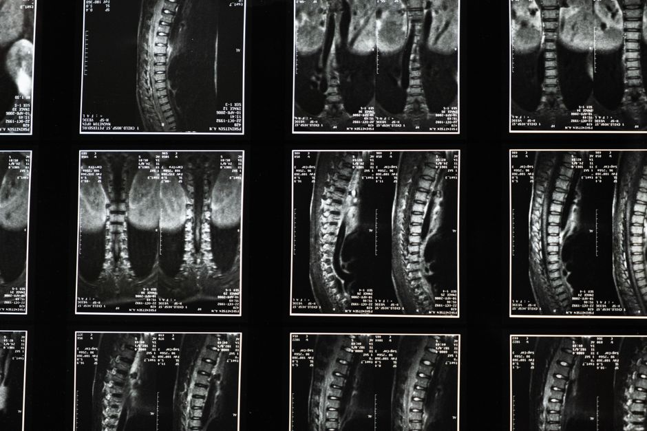
A CT scan measures bone density by sending X-rays through the body from many angles and calculating how much of that radiation is absorbed by bone versus surrounding tissue. Dense bone absorbs more X-rays, so the scanner’s computer can build a three-dimensional picture and estimate the mineral content inside a specific volume of bone. This is different from standard bone density testing, which uses a different type of scan and reports results on a scale most people recognize.
How Does a CT Scan Actually Measure Bone Density?
A CT scanner rotates an X-ray source and detectors around the body. As the beam passes through tissue, some X-rays are absorbed and some pass through to the detectors. Bone absorbs more than fat or muscle because calcium and other minerals in bone are denser.
The scanner repeats this from hundreds of angles. A computer then reconstructs cross-sectional images, called slices, that show the inside of the body in fine detail. From these slices, software can isolate a specific section of bone and calculate how much mineral is present per unit of volume.
The measurement is usually called volumetric bone mineral density, reported in milligrams per cubic centimeter (mg/cm³). Because it is a true three-dimensional measurement, it captures the thickness and internal structure of bone in a way a flat two-dimensional scan cannot.
This is the key difference from DXA, the standard test for osteoporosis. DXA measures bone mineral density in grams per square centimeter (g/cm²) — an area, not a volume. CT measures a volume. That distinction matters for how results are interpreted and which test a doctor chooses.
What Is the Difference Between a CT Scan and a DXA Scan?
DXA (dual-energy X-ray absorptiometry) is the standard test used to diagnose osteoporosis. It is fast, uses a low radiation dose, and reports results as a T-score, which compares your bone density to that of a healthy young adult.
Quantitative CT (QCT) and its newer form, volumetric QCT, measure bone density in three dimensions. They can separate the outer cortical bone from the inner trabecular bone, which DXA cannot do. Trabecular bone is more metabolically active and often changes earlier in disease.
Here is how the two compare on the features that matter most:
| Feature | DXA | CT (QCT) |
|---|---|---|
| Measurement type | Area (g/cm²) | Volume (mg/cm³) |
| Radiation dose | Very low | Higher |
| Reports T-score | Yes | Not directly |
| Separates bone types | No | Yes |
| Common use | Diagnosis and monitoring | Specialized cases |
Because DXA is the test validated for diagnosing osteoporosis, most clinical guidelines are built around it. CT is generally used when DXA results are unclear, when a doctor needs to look at bone structure in more detail, or when a CT is already being done for another reason.
When Would a Doctor Order a CT for Bone Density?
CT is not the first-line test for bone density in most cases. It tends to be ordered in specific situations where its strengths matter.
- When a CT scan is already planned for another purpose, such as evaluating the spine or abdomen, and bone density can be assessed from the same images.
- When DXA results are hard to interpret because of spinal arthritis, prior spine surgery, or calcium deposits that can falsely raise DXA readings.
- When a doctor wants to evaluate trabecular bone specifically, since it can show changes before DXA does.
- In some research and specialty settings where three-dimensional bone structure is the focus.
The higher radiation dose is the main reason CT is not used routinely for screening. A DXA scan exposes a person to a very small amount of radiation, while a CT scan delivers more. That trade-off is why DXA remains the standard for most people.


How Are CT Bone Density Results Interpreted?
CT bone density results are reported as a volumetric measurement in mg/cm³. Unlike DXA, there is no single universal T-score cut-off that applies to every CT scanner and protocol.
Different manufacturers and software packages use different reference values. Some clinical centers convert QCT results into a T-score equivalent so they can be compared to DXA-based thresholds. The World Health Organization defines osteoporosis using DXA T-scores, not CT values.
This is a genuine limitation. A CT number from one machine may not mean the same thing as a number from another. That is why interpretation depends heavily on the specific protocol and reference database being used.
In practice, a doctor looks at the CT value alongside other information: your age, sex, medical history, fracture history, and other risk factors. No single number tells the whole story.
What Are the Limits and Risks of CT Bone Density Measurement?
The main drawback is radiation exposure. CT uses more ionizing radiation than DXA. The exact dose depends on the scanner, the body part scanned, and the settings used. For this reason, CT is not recommended as a routine screening tool for osteoporosis in people without specific reasons to need it.
Cost and access are also factors. CT scanners are widely available, but a dedicated QCT bone density study may not be covered by insurance the way DXA often is. Availability of the specialized software varies by facility.
There is also the matter of standardization. Because CT bone density values are not reported on a single universal scale, comparing results across different machines or over time requires care. A change in your number could reflect a real change in bone or simply a different scanner or protocol.
None of this makes CT a bad test. It makes it a specialized one. Used in the right situation, it provides information DXA cannot.
Can CT Replace DXA for Diagnosing Osteoporosis?
No. DXA remains the standard test for diagnosing osteoporosis, and the World Health Organization’s diagnostic criteria are based on DXA T-scores. A T-score of -2.5 or lower at the hip or spine is the established threshold for osteoporosis.
CT can provide useful additional information, especially about bone structure and trabecular bone. Some studies suggest that QCT can predict fracture risk, and in some cases it may identify people at risk that DXA would miss. But it has not replaced DXA as the diagnostic standard.
If you are being evaluated for bone health, the conversation usually starts with DXA. CT comes into play when there is a specific reason to look deeper.
What Should You Know Before Getting a CT for Bone Density?
If your doctor suggests a CT scan to assess bone density, ask why. In many cases, there is a clear reason — a DXA result that was hard to read, a need to look at bone structure, or a CT already planned for another purpose.
Ask about radiation dose and whether a DXA would give the information needed with less exposure. Ask how the results will be interpreted and what reference values the facility uses.
Bone health is not just about one scan. Factors like calcium intake, vitamin D status, physical activity, smoking, and certain medications all affect bone over time. A scan is one piece of a larger picture.
If you have concerns about your bone health, talk with your doctor about which test is right for your situation. The answer depends on your history, your symptoms, and what information is needed.
Frequently Asked Questions
How does a CT scan measure bone density?
A CT scan measures bone density by passing X-rays through the body from many angles and calculating how much radiation is absorbed by bone. Dense bone absorbs more X-rays, allowing the computer to estimate mineral content per unit of volume.
Is a CT scan better than a DXA scan for bone density?
Neither is universally better. DXA is the standard test for diagnosing osteoporosis and uses much less radiation, while CT provides three-dimensional detail and can separate cortical from trabecular bone.
What radiation dose does a CT bone density scan use?
A CT bone density scan uses more radiation than a DXA scan, though the exact dose depends on the scanner and protocol. This higher dose is why CT is not used for routine osteoporosis screening.
Can a CT scan diagnose osteoporosis?
Osteoporosis is diagnosed using DXA T-scores, not CT values. CT can provide additional information about bone structure, but it has not replaced DXA as the diagnostic standard.
