
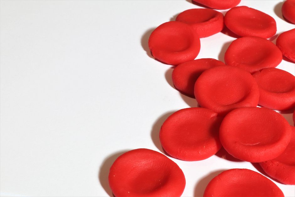
Cell division in the small intestine happens in the crypts of Lieberkühn, which are small pits at the base of the villi. These crypts are the production site for new intestinal cells. The cells move upward from the crypt to the tip of the villus, where they are shed. This entire journey takes about 3 to 5 days in a healthy adult.
What Are Villus Crypts?
The lining of your small intestine is not flat. It is covered with millions of tiny, finger-like projections called villi. Each villus increases the surface area of your gut, which helps your body absorb nutrients from food.
At the base of each villus, there is a small indentation called a crypt. These are the intestinal glands, also known as the crypts of Lieberkühn. They sit just below the surface of the intestinal lining. The crypts are where the entire lining regenerates itself.
Your intestinal lining faces a harsh environment. Digestive enzymes, stomach acid, and the physical movement of food constantly damage these cells. To keep up, your body replaces the entire lining roughly every week. The crypts make this rapid replacement possible.
Where Does Cell Division Occur In The Villus Crypts?
Cell division occurs at the bottom of the crypt, in a region called the stem cell zone. These are the intestinal stem cells. They are the only cells in the intestinal lining that divide continuously.
Stem cells in the crypt divide to produce two types of cells. One remains in the crypt as a stem cell. The other becomes a transit-amplifying cell. These transit-amplifying cells divide several more times, increasing the number of cells available to move upward.
This process is tightly controlled. The cells divide only in the lower two-thirds of the crypt. They stop dividing once they reach the upper third. By the time a cell reaches the top of the crypt, it has matured into a functional intestinal cell.
How Do Cells Move From the Crypt to the Villus?
Once cells stop dividing, they do not stay in the crypt. They migrate upward toward the tip of the villus. This movement is driven by pressure from new cells forming below them.
During this migration, the cells change. They develop microvilli, which are tiny hair-like structures on their surface. These microvilli further increase the surface area for nutrient absorption. The cells also produce digestive enzymes that help break down food.
Most of these migrating cells become enterocytes, which are the main absorptive cells of the intestine. Some become goblet cells, which produce mucus. Others become enteroendocrine cells, which release hormones. Each cell type has a specific job, but they all originate from the same stem cells in the crypt.
When the cells reach the tip of the villus, they undergo a process called apoptosis, or programmed cell death. They are then shed into the intestinal lumen and pass out of the body with the stool. This shedding is normal and continuous.


Why Is This Process So Fast?
The intestinal lining has one of the fastest cell turnover rates in the human body. Research consistently shows that the entire lining is replaced every 3 to 5 days. This speed is necessary because the lining takes constant damage.
Food particles scrape against the villi. Digestive enzymes break down proteins, including the proteins in your own cells. Bacteria in the gut produce toxins that can harm the lining. The rapid replacement ensures that damaged cells are removed quickly and replaced with healthy ones.
This fast turnover also protects you from cancer. Because cells divide so rapidly, the system has built-in checkpoints. If a cell develops a harmful mutation, it is usually shed within days. This limits the time a damaged cell has to grow into a tumor.
What Happens When Cell Division Goes Wrong?
Problems arise when cell division in the crypts becomes unbalanced. If the stem cells divide too slowly, the lining thins. This can lead to poor nutrient absorption and a condition called intestinal atrophy.
Celiac disease is a well-known example. In people with celiac disease, gluten triggers an immune reaction that damages the villi. The crypts respond by producing more cells to compensate. This leads to crypt hyperplasia, where the crypts become longer and deeper in an attempt to repair the damage.
If cell division becomes uncontrolled, it can lead to colorectal cancer. Most colorectal cancers begin in the crypts. A series of genetic mutations can cause the stem cells to divide without stopping. Over time, this creates a polyp, which may develop into cancer.
The balance between cell division and cell death is critical. Too little division causes tissue loss. Too much division, or division without proper cell death, can cause cancer. The body maintains this balance through multiple signaling pathways.
What Controls Cell Division in the Crypts?
Several signaling pathways regulate cell division in the crypts. The most important is the Wnt signaling pathway. It keeps stem cells in their undifferentiated state and promotes their division.
When Wnt signaling is too active, cells divide excessively. This is a common feature in colorectal cancer. When Wnt signaling is too weak, the crypts shrink and the lining cannot regenerate properly.
Another important pathway is the Notch signaling pathway. It determines what type of cell a dividing cell becomes. It decides whether a cell becomes an absorptive enterocyte or a secretory cell like a goblet cell. This decision happens within the crypt, before the cell begins its migration.
Growth factors and hormones also influence cell division. The body can increase cell production when needed, such as after intestinal injury. This adaptability helps the intestine recover from damage, but it also creates a risk if the regulation fails.
Can Diet or Lifestyle Affect Crypt Cell Division?
Diet can influence the rate of cell division in the crypts, but the effect is modest. Caloric intake, fiber, and certain nutrients can alter the balance of the gut lining. However, the body tightly controls the baseline rate of division.
Short-chain fatty acids, produced when gut bacteria ferment fiber, are known to support healthy crypt function. They provide energy for colon cells and may help regulate cell division. Some studies suggest that a high-fiber diet reduces the risk of colorectal cancer, but the evidence is not definitive.
Fasting and caloric restriction have been shown to reduce cell division rates in animal studies. This reduction may lower cancer risk, but the effect in humans is not well established. No clinical guidelines currently recommend specific diets to change crypt cell division rates.
Chronic inflammation can increase cell division in the crypts. Conditions like inflammatory bowel disease cause prolonged inflammation, which stimulates the crypts to produce more cells. This increased division raises the risk of mutations and colorectal cancer over time.
Why Understanding Crypt Cell Division Matters
Understanding where and how cell division occurs in the crypts matters for several reasons. It explains how the gut repairs itself after injury or infection. It also helps researchers understand how colorectal cancer begins.
Most colon cancer treatments target rapidly dividing cells. Chemotherapy drugs like 5-fluorouracil work by killing dividing cells. This is why they are effective against cancer, but it is also why they cause gastrointestinal side effects. The drugs damage the healthy dividing cells in the crypts, not just the cancer cells.
Researchers are studying ways to protect the crypt stem cells during chemotherapy. If the stem cells survive treatment, the gut lining can regenerate faster. This could reduce the severity of chemotherapy side effects.
The crypt system is also a model for studying stem cells in general. Because these cells divide rapidly and are easy to observe, they help scientists understand how stem cells behave in other parts of the body. This research may lead to new treatments for conditions involving tissue damage or degeneration.
Frequently Asked Questions
How long does it take for intestinal cells to renew?
The entire intestinal lining is replaced every 3 to 5 days in a healthy adult. Cells divide in the crypt, migrate up the villus, and are shed at the tip.
What type of cells are found in the villus crypts?
The crypts contain stem cells, transit-amplifying cells, and mature cells preparing to migrate. The mature cells include enterocytes, goblet cells, and enteroendocrine cells.
Does cell division stop at the top of the crypt?
Yes, cells stop dividing in the upper third of the crypt. They migrate upward and mature into functional cells without further division.
Can damage to the villus crypts cause disease?
Yes, damage or dysfunction in the crypts can lead to poor absorption, tissue thinning, or uncontrolled growth. Conditions like celiac disease and colorectal cancer involve abnormal crypt behavior.
