
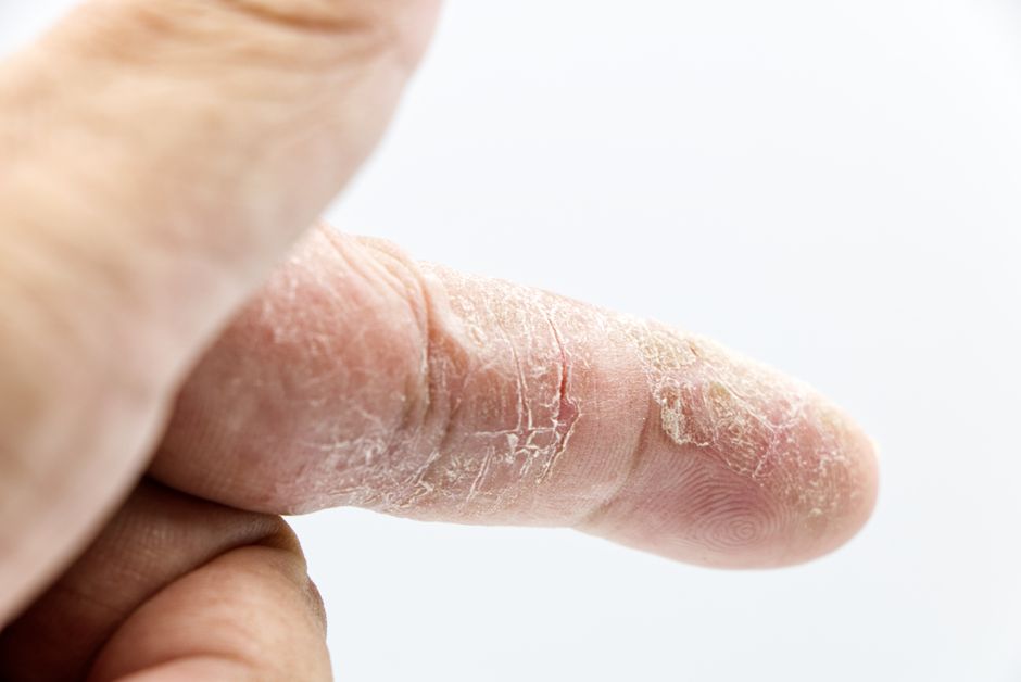
Spongiotic dermatitis is not a disease you catch or a rash with one single cause. It is a pattern that a pathologist sees under a microscope when fluid builds up between the cells of the outer layer of skin. That fluid separates the cells and creates tiny gaps, and the tissue looks like a sponge. Many different skin conditions can produce this pattern, which is why a biopsy report that says “spongiotic dermatitis” is a description of what is happening, not a final diagnosis. The job of your doctor is to figure out which condition is driving it.
What Is Spongiotic Dermatitis And What Causes It?
Spongiosis is the medical term for fluid collecting in the spaces between skin cells in the epidermis, the top layer of your skin. When this happens, the cells pull apart slightly and the tissue takes on a spongy appearance. This is the defining feature of a group of skin conditions that doctors call the spongiotic dermatitides.
The causes are not one thing. They include allergic contact dermatitis, irritant contact dermatitis, atopic dermatitis (eczema), nummular dermatitis, seborrheic dermatitis, stasis dermatitis, and certain drug reactions. Each of these has a different trigger, but they can all produce the same spongiotic pattern when a dermatopathologist looks at a skin sample.
So the phrase on a biopsy report is a starting point, not an answer. It tells your doctor what category of skin problem you have. The next step is matching that pattern to your history, where the rash appears on your body, and what makes it better or worse.
What Does Spongiosis Actually Look Like Under a Microscope?
To understand why spongiotic dermatitis behaves the way it does, it helps to know what is happening at the tissue level. The epidermis is built from cells called keratinocytes, packed tightly together to form a barrier. When inflammation releases fluid into that layer, the fluid pushes between the cells and widens the spaces between them.
Pathologists describe several features that often appear together with spongiosis. These include:
- Intercellular edema — the fluid between cells that defines spongiosis
- Perivascular inflammatory infiltrate — immune cells gathering around small blood vessels in the upper skin
- Exocytosis — immune cells moving into the epidermis itself
- Microvesicles — tiny fluid pockets that can merge into larger blisters
When the fluid pockets grow large enough, they can form visible blisters. This is why some spongiotic conditions, like poison ivy, produce small bumps and blisters while others stay dry and scaly. The amount of fluid and where it collects shapes what you see on the surface of your skin.
One clarifying point: spongiosis is a reaction pattern, not a diagnosis. Two people can have identical biopsy findings and completely different conditions with different triggers and different treatment needs.
Which Skin Conditions Produce Spongiotic Dermatitis?
Several distinct conditions share this microscopic pattern. Knowing the list helps explain why the same biopsy result can mean very different things for different people.
Allergic contact dermatitis happens when your immune system reacts to something touching your skin, such as nickel, poison ivy, or certain preservatives in cosmetics. It usually appears where the skin made contact and often takes a day or two to develop.
Irritant contact dermatitis is different. It does not involve an allergic immune response. It happens when a substance directly damages the skin barrier, like repeated hand washing, harsh soaps, or solvents. This is common in people whose jobs involve frequent exposure to water and chemicals.
Atopic dermatitis, also called eczema, is a chronic condition linked to a weakened skin barrier and an overactive immune response. It tends to run in families with a history of eczema, asthma, or hay fever.
Nummular dermatitis produces coin-shaped, itchy patches, most often on the arms and legs. The exact cause is not fully understood, but dry skin and irritation appear to play a role.
Seborrheic dermatitis affects oily areas like the scalp, face, and upper chest. It is associated with a type of yeast that lives on normal skin, though the yeast is not considered the sole cause.
Stasis dermatitis occurs on the lower legs when blood pools due to poor circulation in the veins. The resulting fluid buildup and inflammation can produce spongiosis.
Drug reactions from certain medications can also trigger a spongiotic pattern, sometimes as part of a broader reaction.





What Are the Symptoms of Spongiotic Dermatitis?
The symptoms depend on which underlying condition is causing the spongiosis, but a few features are common across the group. Itching is the most frequent complaint, and in some conditions it can be intense.
You may notice redness, scaling, small fluid-filled bumps, or blisters. In chronic cases where the skin has been irritated for a long time, the skin can thicken and the normal skin lines become more pronounced, a change doctors call lichenification.
Where the rash appears offers important clues. Contact dermatitis tends to show up where the skin touched the trigger. Atopic dermatitis often affects the inside of the elbows and behind the knees in older children and adults, though it can appear elsewhere. Stasis dermatitis appears on the lower legs. Seborrheic dermatitis favors the scalp, eyebrows, and sides of the nose.
Because the symptoms overlap so much, a physical exam alone is sometimes not enough to tell these conditions apart. That is one reason a doctor may take a small skin sample for testing.
How Is Spongiotic Dermatitis Diagnosed?
Diagnosis usually starts with a careful history and physical exam. Your doctor will ask when the rash started, what it looks like, where it appears, what makes it worse, and whether you have a personal or family history of eczema, asthma, or allergies.
A skin biopsy is the only way to confirm that spongiosis is present. During the procedure, a doctor numbs a small area of skin and removes a sample about the size of a pencil eraser. A pathologist examines it under a microscope and reports the pattern they see.
Patch testing may be used when allergic contact dermatitis is suspected. In this test, small amounts of common allergens are applied to your back under patches and left in place for a set period, then the skin is checked for reactions. This can help identify a specific trigger.
Blood tests and other evaluations may be ordered if a systemic cause or drug reaction is suspected. The exact workup depends on your individual situation, and no single test rules in or out every possible cause.
How Is Spongiotic Dermatitis Treated?
Treatment targets the underlying condition, not the spongiosis itself. Because the causes vary so much, the right approach for one person may be wrong for another.
For contact dermatitis, the most important step is identifying and avoiding the trigger. Once contact stops, the rash often settles on its own, though it can take time. For irritant contact dermatitis, protecting the skin barrier with gentle cleansers and moisturizers is central.
For atopic dermatitis and other inflammatory forms, doctors commonly prescribe topical corticosteroids to reduce inflammation. These are widely used and well established for short-term control of flares. How long and how often to use them depends on the potency, the body area, and your age, so they should be used under medical guidance rather than indefinitely on your own.
Other options your doctor may consider include topical calcineurin inhibitors, which are non-steroid creams sometimes used on sensitive areas like the face, and oral antihistamines for itch. For more severe cases, phototherapy or systemic medications may be considered. The evidence for these treatments varies by condition and severity, and a specialist can help weigh the options.
Moisturizing regularly supports the skin barrier and is a standard part of managing most chronic spongiotic conditions. It is not a cure, but it can reduce how often flares happen for some people.
When Should You See a Doctor?
See a doctor if a rash is spreading, is very itchy or painful, is not improving with basic care, or keeps coming back. You should also seek care if you notice signs of infection, such as increasing warmth, swelling, pus, or fever.
A rash that appears suddenly and covers a large area, or one that comes with swelling of the face or lips or trouble breathing, needs urgent medical attention. These can signal a serious reaction.
Because so many conditions produce spongiotic dermatitis, a clear diagnosis matters. Treating the wrong condition can delay relief, and some triggers, like a medication, need to be identified quickly.
Frequently Asked Questions
Is spongiotic dermatitis contagious?
No, spongiotic dermatitis is not contagious because it is a reaction pattern in the skin, not an infection you can pass to others. The underlying conditions that cause it, such as eczema or contact dermatitis, also do not spread from person to person.
What is the most common cause of spongiotic dermatitis?
There is no single most common cause, because the pattern is shared by several conditions including contact dermatitis, atopic dermatitis, and nummular dermatitis. Which one is most likely depends on your history, the location of the rash, and what triggers it.
Does spongiotic dermatitis go away on its own?
It can improve on its own if the trigger is removed, such as stopping contact with an allergen or irritant. Chronic conditions like atopic dermatitis tend to come and go and usually need ongoing management rather than resolving permanently.
Can a biopsy tell which condition I have?
A biopsy confirms that spongiosis is present but often cannot pinpoint the exact condition by itself. Doctors combine the biopsy findings with your history, symptoms, and sometimes patch testing to reach a diagnosis.
