
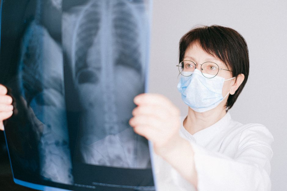
A chest radiograph, commonly called a chest X-ray, is a quick, painless imaging test that uses a small amount of radiation to create pictures of the structures inside your chest. It shows your lungs, heart, blood vessels, airways, and the bones of your spine and chest wall. Doctors use it as a first-line tool to check for many common conditions, from pneumonia and lung cancer to heart failure and rib fractures.
What Is a Chest Radiograph Used to Diagnose?
Chest radiographs are one of the most common medical imaging tests in the world. The CDC reports that millions are performed each year in the United States alone. They are often the first step when someone has a persistent cough, chest pain, shortness of breath, or a fever of unknown origin.
Research shows that a chest X-ray can reliably detect pneumonia, especially when a person has symptoms. A study published in Chest found that chest radiographs have a sensitivity of roughly 70% for diagnosing pneumonia. That means it catches about 7 out of 10 cases. The other 3 may need a CT scan for a clearer view.
Other common diagnoses include:
- Lung cancer – A chest X-ray can spot a mass or nodule, though it often misses smaller early-stage tumors.
- Heart failure – Fluid buildup in the lungs, called pulmonary edema, shows up clearly on an X-ray.
- Pneumothorax – A collapsed lung appears as a dark area where the lung has pulled away from the chest wall.
- Rib fractures – Broken ribs are visible, though hairline fractures can be missed.
- Enlarged heart – The cardiothoracic ratio on the image tells doctors if the heart is bigger than normal.
Some studies suggest that chest X-rays are also used to monitor chronic conditions like COPD and tuberculosis. They are not perfect, but they are fast, cheap, and widely available.
How Does a Chest Radiograph Work?
A chest radiograph works by passing a controlled beam of X-rays through your chest. Different tissues absorb different amounts of radiation. Dense tissues like bone appear white on the image. Air-filled spaces like the lungs appear black. Soft tissues like the heart and muscles appear in shades of gray.
The standard view is called a posterior-anterior (PA) view. You stand facing the detector plate while the X-ray machine is behind you. A second view from the side, called the lateral view, is often taken to give a 3D perspective. Together, these two images help radiologists see the front, back, and sides of your chest.
You will be asked to take a deep breath and hold it for a few seconds. This stops your lungs from moving and gives a sharper image. The entire process takes less than 15 minutes. You feel nothing during the scan.
A key point: the amount of radiation in a chest X-ray is very low. The American College of Radiology states that a standard chest X-ray exposes you to about 0.1 mSv of radiation. That is roughly the same amount you get from natural background radiation over 10 days. It is considered safe for adults.
What Are the Risks of a Chest Radiograph?
The main risk of any X-ray is radiation exposure. However, the dose from a chest radiograph is very low. As mentioned, it is about 0.1 mSv. For context, a CT scan of the chest delivers around 7 mSv — 70 times more. The risk of developing cancer from a single chest X-ray is extremely small. The National Cancer Institute says the benefit of finding a serious condition far outweighs this tiny risk.
There is no evidence that a single chest X-ray causes any immediate side effects. You will not feel anything during or after the scan. Some people worry about the radiation accumulating over multiple scans. That is a valid concern if you need many X-rays over a short period. But for routine use, the risk remains very low.
Pregnant women are a special case. The radiation can harm a developing fetus. If you are pregnant or think you might be, tell your doctor. They may choose a different test, like an ultrasound, or delay the X-ray if it is not urgent.
Some people report feeling anxious or claustrophobic during the scan. This is normal. The machine is large and you have to stay still. But you are not enclosed like in an MRI machine. Most people find it easy.





What Are the Limitations of a Chest Radiograph?
Chest radiographs have real limits. They are good at showing big problems, but they can miss small or subtle ones. A study in Radiology found that chest X-rays miss about 20% of lung cancers that are later found on CT scans. Early-stage lung cancer is especially hard to see because the tumor may be tiny or hidden behind the heart or ribs.
Another limitation is that a chest X-ray cannot tell you what a finding is made of. It can show a spot on your lung, but it cannot tell if that spot is cancer, an infection, or scar tissue. A CT scan or biopsy is needed to find out.
Obesity can also reduce image quality. Extra tissue makes it harder for the X-rays to pass through, leading to a blurrier picture. People who are very large may need a different imaging method.
Some studies suggest that chest X-rays are not reliable for diagnosing pulmonary embolism (a blood clot in the lung). A CT pulmonary angiogram is the standard test for that condition. Chest X-rays are also poor at showing the details of the heart valves or coronary arteries.
Despite these limits, chest radiographs remain a valuable first test. They are quick, cheap, and can rule out many common problems. When something looks suspicious, the doctor will order a more detailed test.
How Do You Prepare for a Chest Radiograph?
Preparation is simple. You do not need to fast or stop any medications. You will be asked to remove any jewelry, glasses, or metal objects from your upper body. Metal can block the X-rays and create artifacts on the image. You will also need to remove your shirt and bra and put on a hospital gown.
Tell the technologist if you are pregnant or have had recent X-rays. They will adjust the procedure if needed. You do not need to bring anything special. The test is done in a radiology department, clinic, or hospital.
During the scan, you will stand against a flat plate. The technologist will position you and step behind a shield. They will ask you to take a deep breath and hold it. Then they press a button. The machine makes a humming or clicking sound. That is it. You can breathe normally after a few seconds.
There is no recovery time. You can go back to your normal activities immediately. The results are usually sent to your doctor within a few hours to a day. Some facilities give you a CD with the images.
What Do the Results of a Chest Radiograph Mean?
Results come as a written report from a radiologist. The report describes what the images show. It will mention the size and shape of your heart, the clarity of your lungs, and the condition of your bones. It will also note any abnormalities.
Common findings and what they may mean:
| Finding on X-ray | What it may indicate |
|---|---|
| White patches in lungs | Pneumonia, fluid, or scar tissue |
| Dark area where lung should be | Collapsed lung (pneumothorax) |
| Enlarged heart shadow | Heart failure or cardiomyopathy |
| Nodule or mass | Tumor, infection, or benign growth |
| Fluid at lung base | Pleural effusion |
| Broken bone line | Rib fracture |
A normal chest X-ray does not mean you are perfectly healthy. It means no obvious abnormalities were seen. Some conditions, like early emphysema or small tumors, can be invisible on X-ray. Your doctor will consider your symptoms and may order more tests if needed.
An abnormal finding does not always mean something serious. Many benign conditions, like old scar tissue from a past infection, look abnormal on X-ray. Your doctor will explain what the finding means in your specific case.
Common Misconceptions About Chest Radiographs
There are several myths about chest X-rays that cause unnecessary worry. One is that the radiation is dangerous. As discussed, the dose is very low. You would need hundreds of chest X-rays in one year to reach a level that raises your cancer risk measurably.
Another myth is that chest X-rays can diagnose all lung problems. They cannot. They miss many small or early-stage conditions. Some people believe a clear X-ray means they do not have lung cancer. That is false. A 2020 study in JAMA Oncology found that 23% of lung cancers were missed on chest X-ray in the year before diagnosis.
Some people think you need to hold your breath for a long time. You only hold it for 2-3 seconds. The X-ray is actually a fraction of a second. The breath hold just keeps your lungs still for that instant.
There is also a belief that chest X-rays are painful. They are not. You may feel cold from the gown or awkward standing still, but there is no pain. The machine never touches you.
Frequently Asked Questions
Frequently Asked Questions
How long does a chest radiograph take?
The entire process takes about 10 to 15 minutes. The actual X-ray exposure is less than one second.
Can a chest radiograph detect heart problems?
Yes, it can show an enlarged heart or fluid in the lungs from heart failure. It does not show the heart’s arteries or valves clearly.
Is a chest radiograph safe during pregnancy?
Doctors avoid it during pregnancy if possible. If it is necessary, the abdomen is shielded to protect the fetus.
Do I need to remove all my clothes for a chest X-ray?
No, you only remove clothing and jewelry from your upper body. You wear a hospital gown for the test.
