
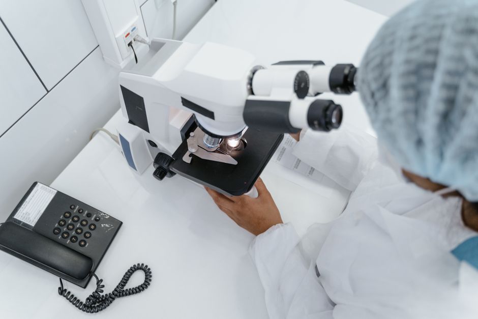
Brightfield microscopy is the most common form of light microscopy used in schools, hospitals, and research labs. It works by shining visible light through a thin specimen, where the light passes directly into the objective lens to create a magnified image. The specimen appears darker than the bright background because dense parts of the sample absorb or scatter light, which is why it is called “brightfield.”
This technique is simple, reliable, and affordable, making it the standard tool for examining stained cells, tissues, and microorganisms. If you have ever looked at a slide in a biology class, you have used brightfield microscopy. While it cannot show fine details inside living cells without stains, it remains the backbone of routine laboratory work.
What Is Brightfield Microscopy And How Does It Work?
Brightfield microscopy uses visible light from a lamp below the stage. The light passes through a condenser, which focuses it onto the specimen. The light then travels through the sample and into the objective lens, which magnifies the image. Finally, the ocular lens (eyepiece) magnifies it further for your eye to see.
The key feature is that the background remains white or bright while the specimen appears dark. This happens because cellular structures absorb and scatter light differently. Stains like hematoxylin and eosin add color and contrast, making details visible that would otherwise be transparent.
Resolution is limited by the wavelength of visible light. A standard brightfield microscope can resolve objects down to about 200 nanometers. That is roughly half the wavelength of green light. Objects smaller than this, such as viruses or individual protein molecules, cannot be seen with this method.
What Are the Main Parts of a Brightfield Microscope?
Understanding the parts helps you know why each component matters. The system works as a coordinated chain, and each piece has a specific role.
- Illuminator: The light source, usually an LED or halogen bulb, sits at the base.
- Condenser: A lens system that focuses light onto the specimen. It has an iris diaphragm to control light intensity and angle.
- Stage: The flat platform holding the glass slide. It moves up and down for focusing and side to side for scanning.
- Objective lenses: Usually 4x, 10x, 40x, and 100x magnification. These are the primary magnifying lenses.
- Ocular lens: The eyepiece, typically 10x magnification. Total magnification equals objective multiplied by ocular.
- Focus knobs: Coarse and fine adjustment knobs move the stage to bring the specimen into sharp focus.
The 100x objective requires immersion oil. Oil has the same refractive index as glass, so it prevents light rays from bending as they pass from the slide to the lens. Without oil, the image blurs and resolution drops significantly.
What Can You See With a Brightfield Microscope?
Brightfield microscopy works best on specimens that are thin enough for light to pass through. Thick samples appear dark and lack detail because light cannot penetrate them.
Typical samples include blood smears, tissue sections, bacteria smears, plant cross-sections, and cultured cells. These are almost always stained before viewing. Stains bind to specific cellular components and absorb certain wavelengths of light, producing contrast.
Common stains include Gram stain for bacteria, Wright stain for blood cells, and hematoxylin and eosin for tissue sections. Fluorescent stains are not used in brightfield because they require a different light source and filter system.
Living cells are difficult to observe with brightfield microscopy. Unstained cells are nearly transparent, so internal structures are invisible. Even if you use a stain, most stains are toxic to living cells. For live-cell imaging, researchers use phase-contrast or differential interference contrast microscopy instead.





What Are the Advantages of Brightfield Microscopy?
The main advantage is simplicity. The instrument is inexpensive compared to other microscope types. It requires minimal training, and samples are easy to prepare.
Brightfield microscopy is highly reliable. The optics are well understood, and the technology has been refined for over a century. Results are consistent between labs, which matters for clinical diagnostics and research reproducibility.
It is also fast. You can prepare a stained slide and examine it within minutes. This makes it ideal for routine screening, such as checking a blood smear for abnormal cells or identifying bacteria in a patient sample.
What Are the Limitations of Brightfield Microscopy?
The biggest limitation is low contrast. Unstained specimens are nearly invisible. Staining solves this but kills cells and can introduce artifacts that were not present in the living sample.
Resolution is capped at about 200 nanometers. You cannot see viruses, ribosomes, or the internal structure of organelles. Electron microscopes are needed for those details.
Depth of field is shallow. Only a thin plane of the specimen is in focus at one time. Thick samples require focusing through multiple planes, which is tedious and can miss structures.
Brightfield cannot visualize structures that do not absorb or scatter light. Transparent organelles like the nucleus and mitochondria remain invisible without stains. The technique also cannot provide information about molecular composition or dynamic processes in living cells.
How Does Brightfield Compare to Other Light Microscopy Techniques?
Several other techniques exist to overcome brightfield limitations. Each has a specific purpose, and the choice depends on the sample and the question being asked.
| Technique | What It Shows | Best For |
|---|---|---|
| Brightfield | Stained structures | Routine clinical and educational use |
| Phase-contrast | Unstained living cells | Observing live cells and internal dynamics |
| Darkfield | Bright objects on dark background | Thin bacteria, spirochetes, and unstained particles |
| Fluorescence | Specific molecules tagged with fluorophores | Localizing proteins, DNA, or organelles |
| Differential interference contrast | 3D-like relief of unstained cells | Detailed morphology of living cells |
Phase-contrast and DIC convert differences in refractive index into visible contrast. This allows you to see living cells without stains. Fluorescence microscopy uses laser light and filters to excite specific molecules, providing high specificity but requiring more expensive equipment.
Darkfield blocks the central light beam and only collects scattered light. This makes tiny objects appear bright against a black background. It is useful for detecting very thin bacteria like Treponema pallidum, which causes syphilis.
What Are Common Applications in Medicine and Research?
In clinical pathology, brightfield microscopy is the standard method for examining biopsy tissues. Pathologists look for abnormal cell shapes, tissue architecture, and signs of cancer under brightfield illumination.
Hematology labs use brightfield to examine blood smears. They count red cells, white cells, and platelets, and look for abnormal cell populations. This helps diagnose anemia, infections, and blood cancers like leukemia.
Microbiology labs use brightfield to identify bacteria and fungi. The Gram stain categorizes bacteria into gram-positive and gram-negative groups, which guides antibiotic selection. Acid-fast staining detects Mycobacterium tuberculosis in sputum samples.
In research, brightfield is used for basic cell counting, assessing cell morphology, and verifying cell culture health. It is often the first step before more advanced imaging techniques are applied.
How Do You Prepare a Sample for Brightfield Microscopy?
Sample preparation directly affects image quality. Poorly prepared slides produce poor images regardless of microscope quality.
For tissue samples, the specimen is fixed in formalin, embedded in paraffin wax, and sliced into thin sections about 5 micrometers thick. The sections are placed on slides and stained.
For blood smears, a drop of blood is spread across the slide in a thin film, air-dried, and stained with Wright or Giemsa stain.
For bacteria, a smear is heat-fixed to the slide and stained using the Gram method. Heat fixation kills the bacteria and adheres them to the glass.
For cultured cells, cells are grown directly on glass coverslips, fixed, and stained. Alternatively, they can be scraped and centrifuged onto slides using a cytocentrifuge.
Each preparation must be thin enough for light to pass through. Thick clumps of cells appear dark and featureless, making diagnosis impossible.
What Are Common Mistakes Beginners Make?
The most common mistake is starting with the 100x objective. Beginners should always start with the 4x or 10x objective to locate the specimen, then move to higher magnification.
Another mistake is using the coarse focus knob at high magnification. The coarse knob moves the stage rapidly and can crash the objective into the slide, damaging both. Use only the fine focus knob at 40x and 100x.
Forgetting immersion oil is another frequent error. The 100x objective produces a blurry image without oil. Conversely, using oil on a 40x lens ruins the lens and requires thorough cleaning.
Insufficient light is also common. Closing the iris diaphragm too much reduces resolution, while opening it fully can wash out contrast. Adjust the diaphragm to balance brightness and contrast.
Finally, many beginners skip cleaning the lenses. Dirty ocular and objective lenses scatter light and reduce image clarity. Use lens paper and approved cleaning solution only.
Is Brightfield Microscopy Still Relevant Today?
Yes. Despite advances in fluorescence and electron microscopy, brightfield remains the most widely used technique in clinical diagnostics and education.
It is the first-line tool for cancer diagnosis, infectious disease identification, and blood cell evaluation. The equipment is affordable, the methods are standardized, and the results are reproducible across laboratories worldwide.
Automated digital pathology systems now scan brightfield slides and analyze them with software. This combines the reliability of traditional staining with the speed and objectivity of computer analysis.
Brightfield microscopy will not replace advanced techniques, and advanced techniques will not replace it. They serve different purposes. For routine, high-volume, cost-effective examination, brightfield remains unmatched.
Frequently Asked Questions
What is the difference between brightfield and darkfield microscopy?
Brightfield uses direct light that passes through the specimen, making the background bright and the specimen dark. Darkfield blocks direct light and only collects scattered light, making the specimen appear bright against a dark background.
Why do brightfield images have low contrast?
Most cells and tissues are nearly transparent and absorb very little light. Stains are required to increase contrast, but they also kill living cells and can alter the sample’s natural appearance.
Can brightfield microscopy see living cells?
Yes, you can see living cells, but internal details are largely invisible because unstained structures are transparent. For live-cell observation with visible internal detail, phase-contrast or DIC microscopy is preferred.
What is the maximum magnification of a brightfield microscope?
Standard brightfield microscopes reach 1000x total magnification using a 100x objective and a 10x eyepiece. Higher magnification does not improve resolution because the limit is set by the wavelength of visible light, not the lenses.
