
A tracer in medical imaging is a substance that doctors can see inside your body using special cameras. It is usually a tiny amount of a safe chemical combined with a radioactive atom or a contrast agent. Once injected, swallowed, or inhaled, the tracer travels through your body and collects in specific organs or tissues. The imaging machine then detects the tracer and creates detailed pictures that show how your body is functioning.
What Is A Tracer How They Work In Medical Imaging?
A tracer works by attaching itself to a molecule your body naturally uses. For example, glucose is a sugar your cells need for energy. A common tracer combines a radioactive form of fluorine with glucose. This creates a substance called FDG. Your body treats FDG like regular glucose. Cells that use more energy, such as cancer cells, take up more FDG. The scanner then shows bright spots where the tracer collects.
The radioactive part of the tracer emits signals that cameras can detect. These signals are called gamma rays. The cameras use these signals to build a three-dimensional map of your body. This map shows not just where things are, but how active they are. This is different from an X-ray or CT scan, which mainly shows structure. Tracers show function.
What Are The Main Types Of Tracers?
There are two main categories of tracers used in medical imaging. The first type is radioactive tracers used in nuclear medicine. The second type is contrast agents used in MRI and CT scans.
Radioactive tracers are the core of PET and SPECT scans. PET stands for Positron Emission Tomography. SPECT stands for Single Photon Emission Computed Tomography. Both use radioactive tracers that emit gamma rays. A PET scanner detects two gamma rays emitted in opposite directions. A SPECT scanner detects single gamma rays from different angles.
Contrast agents are different. They are not radioactive. Instead, they change how tissues appear on a scan. For example, gadolinium is used in MRI scans. It makes blood vessels and certain tissues appear brighter. Iodine-based contrast is used in CT scans. It highlights blood flow and helps doctors see organs more clearly.
How Do Radioactive Tracers Work Step By Step?
The process starts with a patient receiving the tracer. Most often it is given through an IV line in the arm. The tracer then enters the bloodstream and circulates throughout the body. How long this takes depends on the tracer and what part of the body is being studied.
As the tracer moves, it binds to specific receptors or is taken up by specific cells. For a heart scan, the tracer may be absorbed by heart muscle cells. For a bone scan, the tracer collects in areas of new bone growth. The uptake pattern tells the doctor what is normal and what is not.
After the tracer has had time to distribute, the patient lies on a scanning table. The scanner moves over the body and detects the radiation emitted by the tracer. The data is sent to a computer that creates images. The entire scan usually takes between 30 and 60 minutes depending on the type of study.
One important detail is that the radioactive tracer loses its radioactivity over time. This is called the half-life. Different tracers have different half-lives. Some last a few hours. Others last a few days. The half-life is chosen carefully so the tracer stays active long enough for the scan but does not stay in the body longer than needed.




What Are Tracers Used To Diagnose?
Tracers are used to diagnose a wide range of conditions. In cancer care, PET scans with FDG are used to find tumors, see if cancer has spread, and check if treatment is working. Cancer cells are highly active and use more glucose than normal cells, so they appear bright on the scan.
In cardiology, tracers help evaluate blood flow to the heart muscle. A stress test with a tracer can show areas of the heart that are not getting enough blood. This helps doctors identify blockages in the coronary arteries.
In neurology, tracers are used to evaluate brain function. They can help diagnose conditions like Alzheimer’s disease by showing reduced activity in certain brain regions. They can also help locate the source of seizures in people with epilepsy.
In endocrinology, tracers help evaluate the thyroid gland. Radioactive iodine is taken up by the thyroid. A scan can show whether the gland is overactive, underactive, or contains nodules that need further evaluation.
In bone imaging, tracers can detect fractures, infections, or tumors in the bone. They are especially useful for finding small areas of abnormal bone activity that may not show up on regular X-rays.
Are Tracers Safe?
The radiation exposure from a single diagnostic scan is generally low. It is similar to the radiation you might receive from several X-rays. The amount varies by the type of scan and the tracer used.
For most people, the benefit of getting an accurate diagnosis far outweighs the small risk from radiation. However, the risk is not zero. Radiation exposure is cumulative over a lifetime. Doctors usually only order scans with tracers when they are medically necessary.
Pregnant women and nursing mothers need special consideration. Some tracers can cross the placenta or enter breast milk. If you are pregnant or breastfeeding, tell your doctor before any scan involving a tracer. The doctor can decide whether the scan can be delayed or whether a different imaging method is safer.
Allergic reactions are rare but possible, especially with contrast agents. Iodine-based contrast can cause reactions in some people. Gadolinium can cause a serious condition in people with advanced kidney disease. Tell your doctor about any allergies or kidney problems before your scan.
How Do Tracers Compare To Other Imaging Methods?
Each imaging method has strengths and weaknesses. The choice depends on what question the doctor needs to answer.
| Imaging Method | What It Shows | Common Use |
|---|---|---|
| X-ray | Structure of bones and dense tissue | Fractures, chest infections |
| CT scan | Detailed cross-sectional structure | Trauma, tumors, blood clots |
| MRI | Soft tissue detail without radiation | Brain, joints, spinal cord |
| Ultrasound | Real-time images using sound waves | Pregnancy, gallbladder, blood flow |
| PET scan with tracer | Metabolic activity and function | Cancer staging, heart function |
| SPECT scan with tracer | Function and blood flow | Brain disorders, cardiac perfusion |
CT and MRI provide excellent structural detail. They show the size, shape, and location of organs and abnormalities. But they do not tell you how active a tissue is. A tracer scan adds that functional layer. This is why doctors often combine a CT scan with a PET scan. The CT shows where the abnormality is, and the PET shows whether it is active.
What Are The Limitations Of Tracers?
Tracers are powerful tools, but they have limits. A PET scan can show that an area is active, but it cannot always tell you exactly what kind of tissue it is. An infection and a tumor can both appear bright on a PET scan. Additional tests are often needed to make a final diagnosis.
Some tracers are not specific to one type of tissue. They may accumulate in areas of inflammation as well as areas of cancer. This can lead to false positives, where a scan looks abnormal but no cancer is actually present.
Resolution is another limit. PET and SPECT scans do not show as much fine detail as CT or MRI. Small lesions may be missed. This is why doctors rely on multiple imaging methods together rather than a single scan.
Cost and availability are practical concerns. Tracers are expensive to produce. Most require a cyclotron, a large machine that creates radioactive materials. Not every hospital has one. This is why tracer scans are often done at specialized centers.
What Should You Expect During A Tracer Scan?
You will usually be asked to fast for several hours before a PET scan. This is because food can affect how your body handles glucose, which can change the scan results. Your doctor will give you specific instructions before your appointment.
When you arrive, a nurse or technologist will place an IV line in your arm. The tracer is injected through this line. You will then wait in a quiet room while the tracer circulates through your body. This waiting period can last from 30 to 90 minutes depending on the study.
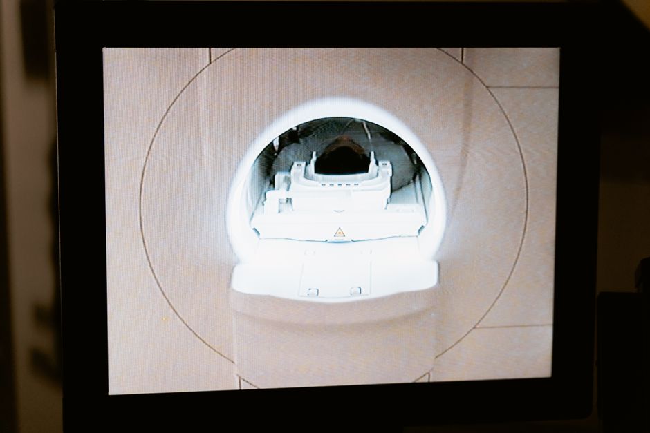
During the scan, you will lie still on a table that slides into the scanner. The scanner is a large machine shaped like a donut. It does not touch you. The scan itself is painless. You may hear humming or clicking sounds from the machine.
After the scan, you can usually go home right away. You should drink plenty of water to help flush the tracer out of your system. The radioactive material will naturally leave your body over the next few hours to a few days, depending on the tracer used.
Frequently Asked Questions
How long does a tracer stay in your body?
Most tracers leave your body within 24 to 48 hours through urine and stool. The exact time depends on the specific tracer and how your kidneys and liver function.
Can you feel the tracer when it is injected?
No, you cannot feel the tracer itself. You may feel a brief cold sensation as the liquid enters your IV, but this is from the fluid temperature, not the tracer.
Is a tracer scan painful?
No, a tracer scan is not painful. The only discomfort is the needle stick for the IV, which lasts a few seconds.
Are tracers safe for children?
Tracer scans are sometimes used in children when medically necessary. The radiation dose is adjusted based on the child’s size, and doctors only order these scans when the diagnostic benefit clearly outweighs the small radiation risk.
