
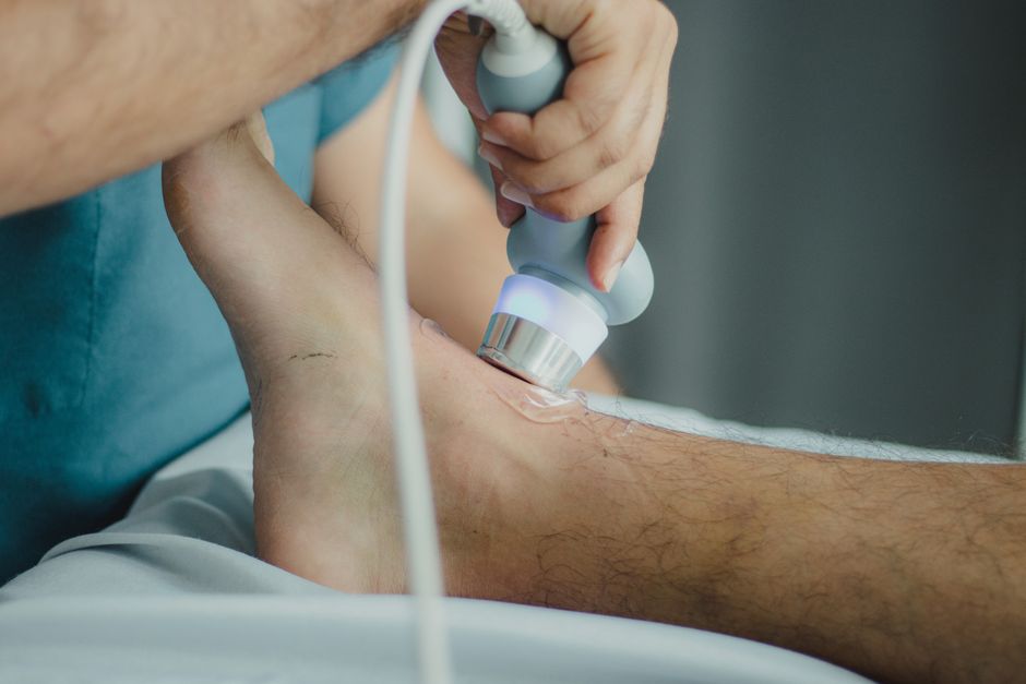
A prostate ultrasound is a medical imaging test that uses sound waves to create pictures of the prostate gland. It helps doctors evaluate the size of the prostate, look for abnormal growths, and guide a biopsy when needed. The test does not use radiation and is generally safe and painless.
What Is a Prostate Ultrasound?
A prostate ultrasound is a type of imaging that produces real-time images of the prostate gland. The medical name is transrectal ultrasound (TRUS) because the most common method involves inserting a small probe into the rectum. The probe sends out sound waves that bounce off the prostate, and a computer turns those echoes into images on a screen.
Doctors use this test to see the shape, size, and texture of the prostate. It is different from an MRI or CT scan because it uses sound instead of magnetic fields or X-rays. The images are detailed enough to spot suspicious areas that may need further testing.
How Does a Prostate Ultrasound Work?
The probe, called a transducer, produces high-frequency sound waves that travel through body tissues. When the sound waves hit the prostate, they bounce back (echo) at different speeds depending on the density of the tissue. The transducer picks up these echoes and sends them to a computer that turns them into a live video image.
During a transrectal ultrasound, the probe is about the size of a finger and is covered with a lubricated condom-like covering. It is gently inserted a few inches into the rectum. Because the rectum sits directly behind the prostate, this approach gives the clearest view. The entire process usually takes 10 to 20 minutes.
Some machines also use Doppler ultrasound to measure blood flow through the prostate. This can help identify areas with increased blood supply, which sometimes indicates inflammation or tumor growth. However, Doppler findings alone do not confirm cancer.
What Is a Prostate Ultrasound Used For?
Doctors order prostate ultrasounds for several reasons. The most common uses include:
- Checking prostate size – Especially in men with symptoms of an enlarged prostate (benign prostatic hyperplasia, or BPH). The test helps measure the size and shape of the gland.
- Guiding a biopsy – When a PSA blood test or digital rectal exam suggests possible cancer, the ultrasound helps the doctor see where to insert the biopsy needle. This is called a transrectal ultrasound‑guided biopsy.
- Evaluating infection – For men with prostatitis (prostate infection), the ultrasound can show swelling or abscesses.
- Monitoring treatment – After procedures like laser therapy or radiation for prostate conditions, ultrasound checks the prostate’s response.
- Investigating infertility – In some cases, ultrasound looks for blockages in the ducts that carry sperm.
It is important to know that a prostate ultrasound alone cannot diagnose prostate cancer. It can show suspicious areas, but only a biopsy can determine whether cells are cancerous.
-
 What Is A Pi Rads 4 Lesion On A Prostate Mri? Key Facts
 -
How to Fix an Enlarged Prostate: Causes, Treatments, and What Actually Works
-
Enlarged Prostate Symptoms Men: Know Everything About BPH
-
Can an Enlarged Prostate Cause Back Pain? What Science Says
-
Prostavive Reviews 2026: Is It Scam Or Legit? [FACTUAL DATA]





Transrectal vs. Transabdominal Ultrasound: What’s the Difference?
Most prostate ultrasounds are done transrectally (through the rectum). A less common method is transabdominal ultrasound, where the probe is placed on the lower belly. The two methods differ significantly in clarity and purpose.
| Feature | Transrectal Ultrasound | Transabdominal Ultrasound |
|---|---|---|
| How it is done | Thin probe inserted into rectum | Probe moved over lower abdomen |
| Image quality | High – shows internal structure of prostate | Lower – less detail, mainly measures size |
| Comfort | Mild pressure, usually well tolerated | Non‑invasive, requires full bladder |
| Common use | Biopsy guidance, cancer evaluation | Quick size check, bladder problems |
A transabdominal ultrasound is sometimes used to measure prostate size in men with BPH, especially when a transrectal exam is not necessary. But for any detailed evaluation, especially when cancer is a concern, transrectal ultrasound is preferred.
How Should I Prepare for a Prostate Ultrasound?
Preparation depends on the type of ultrasound. If you are having a transrectal ultrasound, your doctor may ask you to use an enema or take a laxative the night before or the morning of the test. This clears the rectum so the images are clearer and to reduce the risk of infection if a biopsy is done.
If you are having a transabdominal ultrasound, you will likely need a full bladder. Drinking several glasses of water about an hour before the test and not urinating until after the exam helps push the bladder up for a better view. You should ask your doctor for specific instructions because preparation can vary by clinic.
You should also tell your doctor if you take blood‑thinning medications like warfarin, aspirin, or clopidogrel. These may increase bleeding risk if a biopsy is performed. The doctor may advise you to stop them temporarily.
Are There Any Risks or Side Effects?
Prostate ultrasound is considered very safe. It uses sound waves, not radiation, so there is no exposure to ionizing radiation. The procedure itself rarely causes significant discomfort. Most men describe a feeling of pressure during the probe insertion.
When a biopsy is done along with the ultrasound, the risks increase slightly. Possible side effects include blood in the urine, blood in the semen, or blood in the stool. These usually clear up within a few days. Infection is a rare but more serious risk. Studies estimate that fewer than 1 in 100 men develop a significant infection after a transrectal biopsy. Your doctor may prescribe antibiotics to lower that risk.
What Happens During the Procedure?
You will lie on your side with your knees drawn up toward your chest. The doctor or technician will apply lubricating gel to the probe and gently insert it into the rectum. You may feel a sense of pressure but sharp pain is uncommon. The probe is moved slightly to get different angles. Images appear on a screen in real time.
If a biopsy is needed, the doctor will use the ultrasound images to guide a thin needle through the wall of the rectum and into the prostate. A small device on the probe fires the needle quickly, collecting tiny tissue samples. The sampling takes only a few seconds. After the procedure, you can go home the same day. Most men return to normal activities the next day.
What Do the Results Mean?
A radiologist or urologist reads the ultrasound images and writes a report. For simple size measurements, the report will note the prostate’s volume. A normal adult prostate is roughly the size of a walnut, about 20 to 30 grams. Enlargement is common with age and does not automatically mean cancer.
If the ultrasound shows an abnormal area, the doctor will describe its location, shape, and appearance. But again, the ultrasound cannot classify a spot as cancerous. That requires a biopsy. If you had a biopsy, a pathologist examines the tissue under a microscope and gives a diagnosis – usually a Gleason score that grades how aggressive the cancer looks.
Your doctor will go over the results with you and discuss next steps, whether that is monitoring, treatment, or further tests.
Frequently Asked Questions
Is a prostate ultrasound painful?
Most men feel pressure but not sharp pain during the insertion of the probe. The procedure is usually well tolerated without pain medication.
How long does a prostate ultrasound take?
The imaging part takes about 10 to 20 minutes. If a biopsy is performed, it may add another 5 to 10 minutes.
Do I need a full bladder for a prostate ultrasound?
Only for a transabdominal ultrasound. For a transrectal ultrasound, the bladder is emptied before the exam.
Can a prostate ultrasound detect cancer?
It can show suspicious areas that may be cancer, but a biopsy is the only way to confirm a cancer diagnosis. The ultrasound is used to guide the biopsy needle to those areas.
