
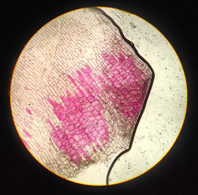
A PAS test is a lab staining method that uses periodic acid and Schiff reagent to detect specific carbohydrates in tissue samples. Pathologists use it to make glycogen, mucins, and certain fungal cell walls visible under a microscope. The stain turns these targets a bright magenta or pink color, which helps identify and diagnose specific diseases.
You may have seen “PAS” on a pathology report and wondered what it means for you. This article explains what the stain does, how it works, and which conditions it helps pathologists identify.
What Is A Pas Test The Histology Stain Explained?
The PAS test is a two-step chemical reaction that colors certain sugars in a tissue sample so they can be seen clearly. “PAS” stands for periodic acid-Schiff, named after the two chemicals involved. It is one of the most common special stains in pathology labs.
Here is the basic chemistry. Periodic acid first reacts with specific carbohydrate structures in the tissue and changes them into a form that can bind to the next chemical. Then the tissue is exposed to Schiff reagent, a colorless solution that turns bright pink or magenta where it attaches. The result is a sharp color contrast that highlights structures a routine stain would miss.
The stain targets what pathologists call glycogen (stored sugar), certain complex sugars called mucins, and the carbohydrate-rich outer layer of some fungi. Normal tissue can contain these substances too, so the pattern and location of the pink color matter as much as its presence.
How Does The PAS Stain Work At The Chemical Level?
The stain works because periodic acid breaks specific chemical bonds in sugar molecules and creates reactive groups that Schiff reagent latches onto. This reaction happens only with certain structures, which is why the stain is selective rather than staining everything.
The key targets are molecules with adjacent sugar units that have a particular chemical arrangement. Glycogen, mucin, and the walls of certain fungi fit this pattern. Most proteins and fats do not, so they stay pale or unstained.
Pathologists often run a control step called diastase digestion. Diastase is an enzyme that breaks down glycogen. If the pink color fades after diastase is applied, the target was likely glycogen. If the color persists, the target is something else, such as a mucin or a fungal wall. This simple comparison helps narrow down what is actually in the tissue.
What Conditions Can A PAS Test Help Diagnose?
The PAS stain helps pathologists evaluate a range of conditions, but it is almost always used alongside other stains and clinical information. It is a supporting tool, not a standalone diagnosis.
- Fungal infections. The cell walls of many fungi contain carbohydrates that PAS stains well. It is commonly used to look for fungal organisms in skin, lung, and other tissue samples.
- Glycogen storage disorders. In rare inherited conditions, abnormal amounts of glycogen build up in organs such as the liver. PAS helps show where glycogen has accumulated.
- Kidney disease. In the glomeruli, the tiny filtering units of the kidney, PAS highlights the basement membranes. Changes in their thickness or appearance can point to certain kidney diseases.
- Liver conditions. PAS can show glycogen in liver cells and help assess certain metabolic and structural liver diseases.
- Mucin-producing cancers. Some tumors produce mucin, and PAS can help confirm its presence. It does not by itself tell whether a tumor is benign or malignant.
- Certain lung and intestinal diseases. PAS is sometimes used to look at mucus-producing cells and specific deposits in these tissues.
The stain is valuable because it is fast, inexpensive, and reliable. But it rarely gives a final answer on its own. A pathologist reads the PAS result together with routine stains, other special stains, and the patient’s symptoms and history.





What Does A Positive PAS Result Actually Mean?
A positive PAS result means the stain found carbohydrates in a particular pattern or location. It does not automatically mean disease. Normal tissues contain glycogen and mucins, so a pathologist interprets the result by where the color appears and how much is there.
For example, a small amount of PAS-positive material in a normal location might be expected. The same amount in an unusual place, or in a pattern that looks abnormal, could point to a problem. The pathologist compares what they see to normal tissue and to the clinical picture.
This is why a PAS report is not something you can read in isolation. Terms like “PAS-positive” describe a staining result, not a diagnosis. The diagnosis comes from the pathologist’s overall interpretation of the sample.
How Is A PAS Test Different From Other Histology Stains?
Different stains highlight different parts of a tissue sample. PAS is one of several special stains, and each has a specific job. Comparing them helps clarify what PAS adds.
| Stain | Main Target | Common Use |
|---|---|---|
| PAS | Glycogen, mucins, some fungal walls | Fungal infections, glycogen storage, kidney and liver disease |
| H&E (hematoxylin and eosin) | General cell and tissue structure | Routine examination of almost all biopsies |
| GMS (Grocott methenamine silver) | Fungal cell walls | Fungal infections |
| Masson trichrome | Collagen and connective tissue | Liver and kidney scarring |
| Alcian blue | Certain mucins | Mucin-producing tissues and tumors |
Pathologists often use PAS and GMS together when looking for fungi, because each can catch organisms the other might miss. The H&E stain is the standard starting point for nearly every biopsy, and special stains like PAS are added when more detail is needed.
What Should You Expect If Your Doctor Orders A PAS Test?
A PAS test is performed on tissue that has already been removed, not on you directly. There is no separate procedure or preparation for the stain itself. It is an extra step a pathologist applies to your biopsy sample in the lab.
If you are having a biopsy, the tissue is removed during that procedure. The lab then processes it, and the pathologist decides whether special stains like PAS would help clarify the diagnosis. The stain adds time to the lab work, usually a small amount compared to the overall processing.
You will not feel anything from the stain. The biopsy procedure itself is the part that involves you, and your care team will explain what to expect for that. Results come back through your doctor, who interprets the pathology report in the context of your symptoms and other tests.
What Are The Limits Of A PAS Test?
PAS is a useful tool, but it is not specific enough to stand alone. Many different substances and tissues can stain positive, so the result must be interpreted carefully. This is a genuine limitation, not a flaw in the test.
A positive result does not confirm a specific disease. It shows that certain carbohydrates are present, and the pattern and context determine what that might mean. Some conditions that look similar under the microscope can produce similar PAS patterns, which is why pathologists rely on multiple stains and clinical information together.
The stain also depends on proper tissue handling. Poorly preserved or incorrectly processed samples can give misleading results. Quality control in the lab matters, and reputable labs run controls to make sure the stain is working correctly.
For some conditions, PAS is helpful but not definitive. Research on the best use of special stains continues, and practices vary between labs. Your pathologist chooses stains based on the specific question at hand.
Frequently Asked Questions
What does PAS stand for in a histology test?
PAS stands for periodic acid-Schiff, named after the two chemicals used in the stain. It detects glycogen, mucins, and certain fungal cell walls by turning them bright pink or magenta.
Is a PAS test the same as a biopsy?
No, a PAS test is a lab stain applied to tissue that was already removed during a biopsy. The biopsy is the procedure that collects the sample; the PAS stain is a step the pathologist uses to examine it.
Does a positive PAS result mean cancer?
No, a positive PAS result does not mean cancer. It simply shows that certain carbohydrates are present, and the pathologist interprets the pattern alongside other stains and your clinical information.
Can a PAS test detect fungal infections?
Yes, PAS is commonly used to help detect fungal organisms because it stains their cell walls well. Pathologists often use it together with other fungal stains to improve detection.
