
A junctional escape rhythm is the heart’s backup system kicking in when its main pacemaker fails or slows down. It originates in the atrioventricular (AV) junction, a backup pacemaker that fires at a slower rate than the heart’s natural pacemaker. On an ECG, this rhythm appears as a slow, regular heartbeat without normal P waves, or with P waves that are inverted or occur after the QRS complex. The root causes are typically conditions that suppress the sinus node, block signals from reaching the ventricles, or increase vagal tone.
What Exactly Is a Junctional Escape Rhythm?
Your heart has a built-in electrical system with a hierarchy of pacemakers. The sinus node in the upper right chamber is the primary pacemaker. It normally fires 60 to 100 times per minute. When it works properly, it suppresses all lower pacemakers.
The AV junction is the next pacemaker in line. It sits between the atria and ventricles. It fires at a slower rate — typically 40 to 60 beats per minute. When the sinus node slows down, fails, or its signal gets blocked, the AV junction steps in. This is a protective mechanism. It keeps blood flowing to your brain and organs even when the main system fails.
Junctional escape rhythm is not a disease itself. It is a symptom of an underlying problem. Think of it as a warning light on your dashboard. The light tells you something needs attention, but the light itself is not the problem.
What Are the Root Causes of a Junctional Escape Rhythm?
The AV junction activates when it stops receiving signals from above. This happens in three main scenarios.
Sinus node dysfunction is the most common cause. The sinus node becomes diseased or damaged and fires too slowly. This condition is often called sick sinus syndrome. It becomes more common with age. Scarring from heart surgery, ischemia, or inflammation can also damage the sinus node.
AV block is the second major cause. The electrical signal leaves the sinus node but gets blocked before it reaches the ventricles. In third-degree heart block, no signals get through at all. The ventricles would stop entirely without the junctional escape rhythm. This is why the escape rhythm is life-saving in complete heart block.
Increased vagal tone is the third cause. The vagus nerve slows the heart during rest and sleep. Athletes often have high vagal tone and resting heart rates below 60. During sleep or deep relaxation, the sinus node may slow enough for the AV junction to fire a few beats. This is usually harmless.
Medications can also trigger junctional escape rhythm. Beta-blockers, calcium channel blockers, and digoxin all slow the heart. In high doses, they can suppress the sinus node enough for the junction to take over.
How Does a Junctional Escape Rhythm Appear on an ECG?
The ECG is the definitive tool for identifying this rhythm. Three features stand out.
Rate is the first clue. The ventricular rate is typically 40 to 60 beats per minute. This is slower than normal sinus rhythm but faster than a ventricular escape rhythm, which fires at 20 to 40 beats per minute.
P waves are the second clue. In normal sinus rhythm, each P wave precedes the QRS complex. In junctional escape rhythm, the P waves are often absent entirely. The signal originates in the AV junction, so it does not depolarize the atria in the normal direction.
When P waves do appear, they are often inverted. The signal travels backward through the atria instead of forward. These inverted P waves may appear before the QRS, hidden within it, or after it. The position depends on where exactly the signal originates and how fast it travels.
QRS complex is the third clue. The QRS is narrow, usually less than 120 milliseconds. This tells you the signal is traveling down the normal conduction pathway. A wide QRS would suggest the escape is coming from the ventricles instead.





What Do the Different Types of Junctional Rhythms Look Like?
Junctional rhythms are classified by their relationship to the sinus node. The ECG appearance differs based on which rhythm is dominating.
Junctional escape rhythm occurs when the sinus node is slow or blocked. The junction takes over completely. The heart rate is 40 to 60. This is the pure form of the rhythm.
Junctional escape beats are single beats that interrupt an otherwise normal rhythm. The sinus node slows momentarily, one junctional beat fires, then sinus rhythm resumes. This is common and often harmless.
Accelerated junctional rhythm occurs when the junction fires faster than its normal rate. The rate is 60 to 100 beats per minute. This happens when the junction becomes irritable rather than when the sinus node fails. Causes include digoxin toxicity, myocardial ischemia, and electrolyte imbalances.
Junctional tachycardia is the fastest form. The rate exceeds 100 beats per minute. This is much less common and usually indicates a significant underlying problem.
Is a Junctional Escape Rhythm Dangerous?
It depends entirely on the context. The rhythm itself is not dangerous. In fact, it is often protective. The danger comes from what caused it.
In a healthy young athlete with high vagal tone, a few junctional escape beats during sleep are benign. No treatment is needed. The same rhythm in an elderly patient with complete heart block is a medical emergency. The junctional rhythm may be the only thing keeping that patient alive, but it is not reliable long-term.
Symptoms matter more than the ECG appearance. Many people feel nothing at all. Others experience fatigue, lightheadedness, or shortness of breath. If the rate drops too low, fainting can occur. The brain needs a minimum heart rate to maintain adequate blood flow. When the escape rhythm cannot keep up with demand, symptoms appear.
Sinus node dysfunction and high-grade AV block often progress over time. A patient with occasional junctional escape beats today may develop complete heart block in the future. This is why any junctional rhythm in a symptomatic patient deserves thorough evaluation.
When Is Treatment Needed for a Junctional Escape Rhythm?
Treatment targets the underlying cause, not the rhythm itself. There is no medication that specifically suppresses junctional escape rhythm, and there should not be. Suppressing it without fixing the cause could stop the heart entirely.
If medications are causing the rhythm, the dose may need adjustment. Digoxin toxicity requires specific management. Beta-blockers or calcium channel blockers may be reduced or changed. This decision should always be made by a physician. Never stop a heart medication on your own.
If the cause is sinus node dysfunction or AV block, a pacemaker may be necessary. Pacemakers are highly effective for these conditions. They restore a normal heart rate and relieve symptoms. The decision to implant a pacemaker depends on symptom severity and the underlying rhythm disorder.
Asymptomatic patients with junctional escape rhythm often need no treatment at all. Close monitoring may be sufficient. The key is identifying why the rhythm is occurring and whether it is likely to progress.
How Is a Junctional Escape Rhythm Diagnosed?
A standard 12-lead ECG is usually sufficient for diagnosis. The characteristic findings appear clearly when the rhythm is active. But junctional rhythms can be intermittent. A single ECG may miss them.
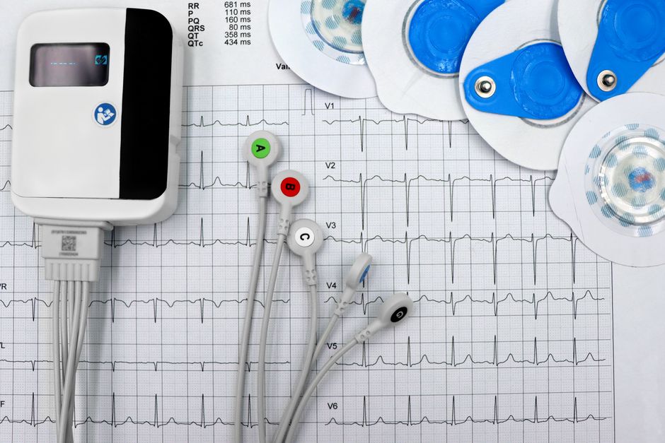
Holter monitoring is used when symptoms are intermittent. This device records the heart rhythm continuously for 24 to 48 hours. Patients keep a diary of symptoms so the doctor can correlate symptoms with rhythm changes.
Event monitors are used for longer periods. These devices are worn for weeks or months. The patient activates the monitor when symptoms occur. This captures rhythms that happen only occasionally.
An electrophysiology study may be needed in complex cases. This involves threading catheters into the heart to map its electrical system. It is invasive but provides detailed information that surface ECGs cannot.
What Is the Difference Between Junctional and Ventricular Escape Rhythms?
This distinction matters because the two rhythms have different implications. Both are backup pacemakers, but they sit at different levels of the conduction system.
Junctional escape rhythm originates in the AV junction. The QRS complex is narrow because the signal travels down the normal His-Purkinje pathway. The rate is 40 to 60 beats per minute. It is relatively stable and reliable.
Ventricular escape rhythm originates in the ventricles themselves. The QRS complex is wide and bizarre because the signal spreads from muscle cell to muscle cell rather than using the fast conduction pathway. The rate is 20 to 40 beats per minute. This rhythm is less reliable and often indicates severe heart disease.
A ventricular escape rhythm is more concerning. It suggests that both the sinus node and the AV junction have failed. This is a medical emergency that usually requires immediate pacing.
Frequently Asked Questions
Can a junctional escape rhythm go away on its own?
Yes, if the underlying cause is temporary. Once the triggering medication wears off or the vagal tone returns to normal, the sinus node usually resumes its role. Persistent junctional rhythm requires investigation of the underlying cause.
Is junctional escape rhythm the same as a heart attack?
No. A junctional escape rhythm is an electrical finding, not a blocked artery. However, a heart attack can damage the sinus node or conduction system and trigger a junctional rhythm as a secondary effect.
What heart rate is considered junctional escape rhythm?
The typical rate is 40 to 60 beats per minute. Rates above 60 are called accelerated junctional rhythm. Rates below 40 suggest the escape is coming from the ventricles instead.
Can anxiety cause a junctional escape rhythm?
Anxiety itself does not directly cause this rhythm. Anxiety increases heart rate through sympathetic activation, which suppresses escape rhythms. However, anxiety can cause hyperventilation and other changes that indirectly affect heart rhythm in susceptible individuals.
