
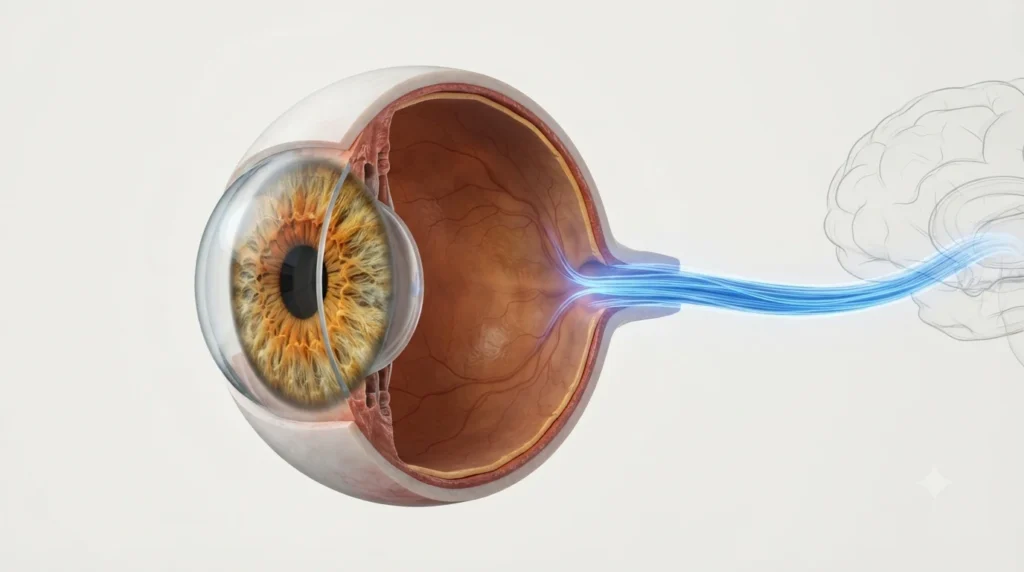
Your eyes do not actually see. They collect light. The optic nerve is what takes that light information and sends it to the brain, where seeing actually happens. It is a cable made of roughly one million nerve fibers, running from the back of each eye straight into the brain. Damage to even a portion of it and your vision changes — permanently. That is how critical this nerve is.
The Job of the Optic Nerve
The optic nerve carries visual signals from your eye to your brain. The retina at the back of your eye captures light and converts it into electrical signals. The optic nerve picks up those signals and delivers them to the visual cortex — the part of the brain that processes what you see.
It handles everything your eyes pick up: brightness, color, contrast, shape, motion. None of that information reaches the brain without the optic nerve doing its job.
Where It Is and What It Looks Like
The optic nerve starts at the back wall of the eyeball and exits through a small opening in the skull called the optic canal. From there, it travels into the brain.
A few things to know about its structure:
- It is made of about one million nerve fibers bundled tightly together
- Each fiber is coated in myelin — a fatty sheath that speeds up signal transmission
- It is technically part of the brain, not a peripheral nerve like those in your hands or feet
- It is the second of the twelve cranial nerves, which is why doctors label it CN II
- An eye doctor can see the front end of it directly through your pupil during a standard eye exam — no surgery needed
That last point matters. The optic nerve is the only part of the central nervous system visible from outside the body. It gives doctors a direct window into brain health, not just eye health.
How Visual Signals Actually Travel
Light enters the eye and hits the retina. Specialized cells in the retina — called photoreceptors — react to that light and pass signals to another layer of cells called ganglion cells. The axons of those ganglion cells are the nerve fibers that make up the optic nerve.
Those fibers gather at a single exit point at the back of the eye, form the optic nerve, and travel through the eye socket into the skull. Inside the skull, they reach a crossing point, then continue to the brain’s visual processing center.
The whole journey — from light hitting your retina to your brain forming an image — happens in milliseconds.
It Does More Than Carry Visual Signals
Most people assume the optic nerve is purely about sight. It is not. It also runs two important reflexes:
- The pupillary light reflex — point a torch at your eye, and your pupil shrinks. That reaction is triggered through the optic nerve. Doctors test this reflex in almost every neurological and eye examination because a sluggish or absent response points to optic nerve damage or brain injury.
- The accommodation reflex — when you shift your gaze from a distant wall to a book in your hands, your lens changes shape to focus. The optic nerve plays a role in triggering that automatic adjustment.
Both reflexes happen without any conscious thought. The optic nerve handles them entirely on its own.
The Optic Disc and Why You Have a Blind Spot
Where the optic nerve exits the back of the eye, there is a small circular patch called the optic disc. No light-sensitive cells exist at this exact spot — the nerve fibers take up all the space. This creates a blind spot in your visual field.
Every human has one. You just never notice it because the brain fills in the gap automatically, using surrounding visual information and input from your other eye.
The optic disc is what your eye doctor examines when they look into your eye with a bright light. A healthy disc has a defined shape and pink-orange color. If it looks pale, swollen, or has an unusually large central depression, something is wrong — either inside the eye or inside the skull.
How the Two Optic Nerves Share the Work
You have one optic nerve per eye. The two meet inside the skull at a crossing point called the optic chiasm.
At this junction, nerve fibers do not simply continue straight to the same side brain. Fibers from the left side of each retina go to the left brain. Fibers from the right side go to the right brain. It is a partial crossing, not a complete one.
This is why damage to one side of the brain can knock out vision on the opposite side — a stroke on the right side of the brain can cause vision loss in the left visual field of both eyes.
The optic chiasm sits directly below the pituitary gland. When a pituitary tumor grows, one of the first things it can press on is the chiasm. The classic symptom is tunnel vision — loss of peripheral sight in both eyes at once — which is often what finally sends someone to a doctor.
Signs Something Is Wrong With the Optic Nerve
Symptoms vary depending on where the damage is and how severe it is. Common warning signs include:
- Blurred or dimmed vision that does not improve with glasses
- Missing patches in your visual field — areas where nothing registers
- Washed-out colors — reds look dull, contrast drops noticeably
- Pain around or behind the eye, especially when moving the eye
- Vision that is worse in bright light than in dim light
- Pupils that react differently to the same light source
Gradual loss is the most dangerous because the brain quietly compensates. Many people with significant optic nerve damage do not realize it until half their vision is already gone.
What Can Damage the Optic Nerve
- Glaucoma is the leading cause worldwide. Raised pressure inside the eye slowly crushes the nerve fibers from the front. It causes no pain and no early symptoms in most cases. By the time vision changes are noticed, significant permanent damage has already occurred.
- Optic neuritis is inflammation of the optic nerve — often linked to multiple sclerosis. It typically affects one eye, comes on over days, and causes pain on eye movement along with sudden vision loss. Many people recover most of their vision, but the nerve is left slightly weakened.
- Reduced blood flow to the nerve, known as ischemic optic neuropathy, cuts off the oxygen supply. Without blood, nerve tissue dies quickly. This is more common in people with high blood pressure, diabetes, or high cholesterol, and usually strikes overnight — many people wake up with vision already gone in one eye.
- Trauma — a direct blow to the eye or skull — can sever or bruise the nerve. Even if the eye itself is undamaged, the nerve behind it can be.
- Toxic substances are an underappreciated cause. Methanol, found in some industrial solvents and counterfeit alcohol, is particularly destructive. Even a small dose can rapidly destroy both optic nerves and cause permanent blindness.
- Vitamin B12 deficiency is another overlooked cause. Prolonged deficiency slowly damages nerve fibers. Vegans, older adults, and people with absorption problems are most at risk. If caught early, stopping the damage is possible.
Can the Optic Nerve Heal
This is the part most people find surprising — and sobering.
No, it cannot regenerate. The optic nerve is part of the central nervous system. Central nervous system tissue does not regrow after damage the way peripheral nerves do. A damaged nerve fiber in your foot can slowly regrow. An optic nerve fiber cannot.
Whatever vision is lost to optic nerve damage stays lost. No surgery, medication, or therapy currently approved for humans can restore dead optic nerve fibers.
This is why the word “early” appears in every piece of advice about eye health. The only real option is to catch damage before it starts or catch it early enough that most fibers are still alive — then slow or stop whatever is causing the harm.
Research is ongoing. Gene therapy, stem cell treatment, and neuroprotective drugs have shown early promise in lab settings. Human trials are years away from producing anything usable.
How to Keep Your Optic Nerve Healthy
You cannot feel the optic nerve failing. That makes proactive care the only real strategy.
- Annual eye exams after 40 — routine pressure checks catch glaucoma before symptoms appear. Family history of glaucoma means starting earlier.
- Control blood pressure and blood sugar — both conditions reduce blood flow to the nerve over time. Keeping them in a healthy range is direct protection.
- Get enough vitamin B12 — especially if you eat little or no meat, or are over 60. A simple blood test can check your levels.
- Never drink home-brewed or unverified alcohol — methanol contamination is a real risk and causes rapid irreversible blindness.
- Treat eye injuries seriously — a blow to the head or eye that causes even brief vision disturbance needs medical evaluation the same day.
- Do not ignore gradual vision changes — it is easy to assume fading vision is just ageing. Get it checked. Glaucoma and other optic nerve conditions feel exactly like that until it is too late.
Frequently Asked Questions
Can you function normally with a damaged optic nerve?
It depends on how much damage has occurred. Partial damage to one optic nerve often allows functional vision, since the other eye compensates. Extensive bilateral damage can cause severe vision loss that affects daily tasks including driving.
Is the optic nerve the same as the retina?
No. The retina detects light and converts it into electrical signals. The optic nerve carries those signals to the brain. They are separate structures with separate roles.
Can a swollen optic nerve return to normal?
Sometimes. Swelling from optic neuritis often reduces with steroid treatment and vision can recover significantly. Swelling caused by ongoing pressure from a tumor or raised brain pressure requires treating the underlying cause first.
Why can a brain problem cause vision loss?
Because the optic nerve connects directly to the brain, and visual fibers travel deep into brain tissue. A stroke, tumor, or pressure buildup in the skull can disrupt those pathways and cause vision loss even when the eyes themselves are perfectly healthy.
Is the optic nerve checked during a regular eye exam?
Yes. The front of the optic nerve — the optic disc — is visible through the pupil. Eye doctors examine its color, size, and shape as part of a standard exam. Changes can signal glaucoma, raised brain pressure, or other serious conditions before symptoms appear.
At what age should you start worrying about optic nerve health?
Eye pressure checks for glaucoma risk should begin at 40, or earlier with a family history. Optic neuritis can affect people as young as 20. Blood flow problems tend to appear after 50. The practical answer: regular eye exams from early adulthood matter regardless of age.
References
- Optic Nerve: Anatomy, Function and Conditions — All About Vision (allaboutvision.com)
- Optic Atrophy: Symptoms, Causes and Treatment — Cleveland Clinic (clevelandclinic.org)
- The Optic Nerve — Anatomy and Function — KenHub (kenhub.com)
- The Optic Nerve (CN II) — Course and Sensory Functions — TeachMeAnatomy (teachmeanatomy.info)
