
An abnormal optic nerve shape almost always comes down to one of two things: pressure inside the eye pushing the optic nerve head forward, or pressure inside the skull pushing it backward. The optic nerve is the cable that carries visual signals from the eye to the brain. When the shape of the optic disc — the visible front end of that nerve — changes, it usually means something is physically pressing on it, or the nerve tissue itself is swollen.
Doctors see this during a routine eye exam with an ophthalmoscope. The optic disc may look raised, cupped, tilted, or blurred. Each pattern points toward different causes, and some are harmless while others need urgent attention.
What Causes an Abnormal Optic Nerve Shape?
The most common cause of a raised, blurred optic disc is swelling from increased pressure inside the skull, a condition called papilledema. The optic nerve is surrounded by cerebrospinal fluid, the same fluid that bathes the brain. When that fluid pressure rises, it pushes against the back of the eye and the optic nerve head swells.
Papilledema is a sign, not a disease itself. It can result from brain tumors, bleeding in the brain, inflammation of the brain or its coverings, blood clots in the veins that drain the brain, or a condition called idiopathic intracranial hypertension where pressure rises without a clear cause. Idiopathic intracranial hypertension is more common in women of childbearing age who carry extra weight, though it can occur in anyone.
A second major cause is glaucoma, where the optic disc becomes cupped — hollowed out — as nerve fibers gradually die. In open-angle glaucoma, this happens slowly over years, often with no symptoms until vision loss is advanced. The pressure inside the eye is frequently elevated, though some people develop glaucoma with normal eye pressure.
A third category involves structural variations that are present from birth. Some people simply have optic discs that are tilted, small, or shaped differently. These are usually harmless and stable over time. The challenge for eye doctors is telling these apart from something dangerous.
How Does Pressure Damage the Optic Nerve?
The optic nerve carries about 1.2 million nerve fibers from the retina to the brain. These fibers are delicate. When pressure rises around them — whether from inside the eye or inside the skull — blood flow to the nerve can be reduced and the fibers can swell or die.
In papilledema, the swelling is usually on both sides. The increased cerebrospinal fluid pressure is transmitted along the optic nerve sheath, which is a tube of tissue surrounding the nerve. Think of it like a balloon inflating around the nerve. The part of the nerve closest to the eye gets squeezed first.
In glaucoma, the damage happens at the optic disc itself. The fibers that carry peripheral vision are often affected first. That is why people with glaucoma can lose side vision without noticing until a lot of damage has occurred. The central vision tends to be spared until later stages.
One non-obvious point: the optic nerve has no pain fibers. You cannot feel pressure building up inside the eye or around the nerve. This is why regular eye exams matter. The nerve can be under strain for a long time before you notice anything.
What Does an Abnormal Optic Nerve Shape Look Like?
Eye doctors describe the optic disc by its color, margins, and the size of the cup in the center. A normal disc is pinkish-orange with clear edges and a small central depression.
When something is wrong, the appearance changes in specific ways:
- Blurred or raised margins — the edges of the disc look fuzzy instead of sharp. This suggests swelling.
- Enlarged cup — the central hollow area gets bigger as nerve fibers die. This is the hallmark of glaucoma.
- Pale color — a white or pale disc can mean the nerve is not getting enough blood or has already lost fibers.
- Tilted disc — the disc sits at an angle. This is often a normal variant but can sometimes be linked to nearsightedness.
- Hemorrhages or spots — small bleeds on or near the disc can occur with swelling or sudden pressure changes.
None of these findings alone confirms a diagnosis. Doctors use additional tests like optical coherence tomography, which measures the thickness of the nerve fiber layer, and visual field testing to see how well the nerve is functioning.





Can a Normal Optic Nerve Look Abnormal?
Yes, and this is one of the trickiest parts of eye care. Some people have optic discs that look concerning but are completely healthy. These are called pseudopapilledema — literally “false swelling.”
Small, crowded optic discs with little room for the normal central cup can look raised even when nothing is wrong. This is more common in people who are farsighted. Drusen — tiny deposits of calcium and protein inside the disc — can also make the disc look swollen. Optic nerve drusen are present in a small percentage of the population and are usually harmless, though they can sometimes affect peripheral vision.
The key difference is stability. True swelling changes over days to weeks. Pseudopapilledema stays the same. That is why doctors often repeat exams or take photographs to compare over time.
When Should You Be Concerned About Optic Nerve Shape?
An abnormal optic disc is not something you can self-diagnose. It is found during an eye exam. What matters is whether it is new, whether it is changing, and whether it is affecting vision or other functions.
Signs that warrant prompt medical attention include:
- Sudden vision changes in one or both eyes
- Headaches that are worse in the morning or when lying down
- Brief episodes of vision loss or dimming, especially with changes in position
- Double vision
- Ringing in the ears that pulses with your heartbeat
- Nausea or vomiting without another explanation
These symptoms, combined with a swollen optic disc, raise concern for raised intracranial pressure. That needs evaluation by a neurologist or neuro-ophthalmologist, often with brain imaging and a spinal tap to measure cerebrospinal fluid pressure.
If the disc is cupped and you have risk factors for glaucoma — age over 60, family history, African American or Hispanic ancestry, diabetes, or high eye pressure — you should have a full glaucoma workup. Glaucoma cannot be cured, but treatment can slow or stop further vision loss. The earlier it is caught, the more vision can be preserved.
What Happens During an Eye Exam for Optic Nerve Shape?
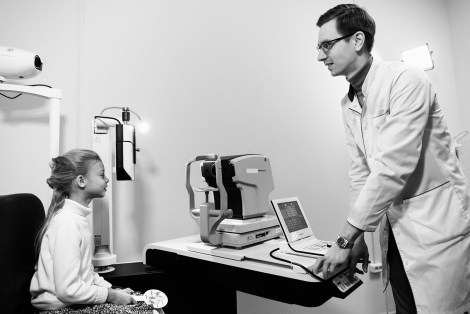
A comprehensive eye exam includes a close look at the optic nerve. The doctor dilates your pupils with drops so the back of the eye can be seen clearly. A slit lamp with a special lens gives a magnified view of the disc.
If the disc looks abnormal, the next steps may include:
- Optical coherence tomography (OCT) — a scan that measures nerve fiber thickness
- Visual field testing — checks for blind spots or peripheral vision loss
- Fundus photography — takes pictures to compare over time
- Fluorescein angiography — uses dye to look at blood flow in the eye, sometimes used to tell true swelling from pseudopapilledema
- Brain imaging — MRI or CT if raised intracranial pressure is suspected
These tests help distinguish between the different causes. They also establish a baseline so changes can be tracked.
What Conditions Are Linked to Abnormal Optic Nerve Shape?
Several conditions can change the shape or appearance of the optic nerve. Some are common and manageable. Others are rare and serious.
Glaucoma is the most common cause of optic nerve cupping. It affects millions of people worldwide and is a leading cause of irreversible blindness.
Papilledema from raised intracranial pressure can be caused by brain tumors, meningitis, encephalitis, cerebral venous sinus thrombosis, or idiopathic intracranial hypertension. The underlying cause determines the treatment.
Optic neuritis — inflammation of the optic nerve — often causes swelling and is frequently linked to multiple sclerosis. It usually comes with pain during eye movement and temporary vision loss.
Ischemic optic neuropathy — reduced blood flow to the nerve — can cause swelling and sudden vision loss. It is more common in older adults and those with cardiovascular risk factors.
Optic nerve hypoplasia — underdevelopment of the nerve — is present from birth and can occur alone or with other brain abnormalities.
Optic nerve drusen — deposits inside the disc — are usually benign but can mimic swelling.
Each of these has different implications and treatments. The appearance of the disc is a starting point, not a final answer.
Can Abnormal Optic Nerve Shape Be Treated?
Treatment depends entirely on the cause. There is no single treatment for an abnormal-looking optic disc.
For papilledema from idiopathic intracranial hypertension, treatment may include weight loss, a medication called acetazolamide that reduces cerebrospinal fluid production, or in severe cases, surgery to relieve pressure. For papilledema from a brain tumor or blood clot, treating the underlying problem is the priority.
For glaucoma, treatment focuses on lowering eye pressure. This can be done with eye drops, laser therapy, or surgery. Lowering pressure does not restore lost vision but can slow or halt further damage. Some people with normal-tension glaucoma may also benefit from pressure-lowering treatment, though the evidence for how low to go is less clear.
For optic neuritis, inflammation often resolves on its own. Corticosteroids may speed recovery but do not appear to change the long-term outcome. For ischemic optic neuropathy, there is no proven treatment that reverses the damage. Managing cardiovascular risk factors is generally recommended.
For pseudopapilledema or optic nerve drusen, no treatment is needed. These are monitored with periodic exams.
Frequently Asked Questions
Can an abnormal optic nerve shape cause blindness?
It can if the underlying cause is not treated. Glaucoma and papilledema can both lead to permanent vision loss if pressure on the nerve continues unchecked.
Is a tilted optic disc serious?
Usually not. A tilted disc is often a normal variant and does not affect vision, though it can sometimes be associated with nearsightedness or visual field changes that need monitoring.
How do doctors tell the difference between true swelling and pseudopapilledema?
They look for changes over time, use imaging like OCT, and sometimes order fluorescein angiography. True swelling changes; pseudopapilledema stays stable.
Can an abnormal optic nerve shape be reversed?
Swelling from raised intracranial pressure can improve once the pressure is treated. Cupping from glaucoma does not reverse, but treatment can prevent further damage.
