
The retina is the light-sensitive tissue at the back of your eye, and it has one of the highest metabolic demands of any tissue in the human body. Retinal blood vessels supply the oxygen and nutrients that tissue needs to convert light into the signals your brain reads as vision. When those vessels narrow, leak, block, or grow abnormally, the retina loses function — and because retinal tissue does not regenerate, that loss can become permanent.
What Are Retinal Blood Vessels and What Do They Do?
Retinal blood vessels are the network of arteries and veins that carry blood into and out of the retina. They branch from the optic nerve head and spread across the inner surface of the retina like rivers across a delta.
Their job is simple and non-negotiable: deliver oxygen and glucose, and carry away metabolic waste. The retina consumes oxygen at a rate comparable to the brain. Interrupt that supply, even briefly, and retinal neurons begin to suffer.
There are two separate blood systems serving the retina:
- Retinal circulation — the vessels visible when a clinician looks into your eye. These supply the inner two-thirds of the retina.
- Choroidal circulation — a dense bed of vessels beneath the retina that supplies the outer layers, including the photoreceptors that actually capture light.
This matters clinically because the retinal vessels are the only blood vessels in the body that can be viewed directly without cutting through skin or tissue. That is why an eye exam can sometimes reveal early signs of systemic disease before a person has any symptoms elsewhere.
How Do Retinal Blood Vessels Affect Your Eye Health?
Healthy retinal vessels maintain a steady, controlled flow of blood. When that control breaks down, the consequences fall into a few broad categories.
Leakage. Damaged vessel walls become permeable. Fluid and blood escape into the retinal tissue, causing swelling. When this happens in the macula — the center of the retina responsible for sharp, detailed vision — it is called macular edema, and it is a leading cause of vision loss in diabetic eye disease.
Blockage. A retinal artery or vein can become occluded. A central retinal artery occlusion cuts off blood supply to most of the retina and is considered a medical emergency. Vein occlusions are more common and typically cause bleeding and swelling that can impair vision over days to weeks.
Abnormal growth. When retinal tissue is starved of oxygen, it releases signals that trigger the growth of new, fragile vessels. This process is called neovascularization. These new vessels are weak and prone to bleeding, and they can lead to scarring and retinal detachment. This is a central mechanism in proliferative diabetic retinopathy.
Narrowing and hardening. Chronic high blood pressure can cause retinal arterioles to narrow and their walls to thicken. Over time this reduces blood flow and can damage the retinal tissue itself. These changes are visible on examination and are graded by clinicians as hypertensive retinopathy.
What Eye Conditions Are Linked to Retinal Blood Vessel Problems?
Several major eye diseases involve retinal blood vessels directly.
Diabetic retinopathy is the most common. Prolonged high blood sugar damages the small vessels in the retina. Early stages may cause no symptoms at all. Over time, it can progress to bleeding, swelling, and abnormal vessel growth. It is a leading cause of blindness in working-age adults in the United States.
Retinal vein occlusion occurs when a vein draining the retina becomes blocked. It can cause sudden or gradual vision loss in one eye. Risk factors include high blood pressure, diabetes, and elevated cholesterol — though not everyone who develops a vein occlusion has these conditions.
Retinal artery occlusion is less common but more urgent. It is essentially a stroke of the eye. Vision loss is typically sudden and painless. It is associated with cardiovascular risk factors and warrants immediate medical evaluation.
Retinopathy of prematurity affects some premature infants. Their retinal vessels are not fully developed at birth, and abnormal vessel growth can occur after delivery. This is why premature infants receive scheduled eye screenings.
Age-related macular degeneration primarily involves the choroidal vessels beneath the retina rather than the retinal vessels on its surface. The “wet” form involves abnormal vessel growth from the choroid. It is a distinct mechanism from diabetic retinopathy, though both involve vascular dysfunction.

What Symptoms Suggest a Retinal Blood Vessel Problem?
Early retinal vessel disease often causes no symptoms. This is not a minor detail — it is the reason regular eye exams matter even when your vision feels fine.
When symptoms do appear, they can include:
- Blurred or distorted vision, especially in the center of your field of view
- Sudden appearance of floaters, sometimes described as a shower of spots
- Flashes of light
- A dark curtain or shadow moving across your vision
- Sudden, painless loss of vision in one eye
- Difficulty seeing at night or in low light
Some of these symptoms overlap with other serious eye conditions, including retinal detachment and vitreous hemorrhage. Any sudden change in vision should be evaluated promptly. Do not wait to see if it improves on its own.
How Are Retinal Blood Vessel Problems Detected?
A dilated eye exam is the starting point. An eye care professional places drops in your eyes to widen the pupils, then examines the retina directly. This allows them to see the vessels, check for bleeding, swelling, or abnormal growth.
Additional testing may include:
- Optical coherence tomography (OCT) — creates cross-sectional images of the retina to detect swelling and thinning.
- Fluorescein angiography — a dye is injected into a vein in your arm, and photographs are taken as it circulates through the retinal vessels. This can reveal leakage and blocked areas.
- Optical coherence tomography angiography (OCTA) — a newer, non-invasive method that maps blood flow in the retina without dye.
Your eye care professional may also recommend blood pressure checks, blood sugar testing, and cholesterol screening if retinal changes suggest an underlying systemic condition.
Can Retinal Blood Vessel Damage Be Prevented or Treated?
Prevention focuses on controlling the conditions that damage blood vessels over time. The evidence here is strong and consistent.
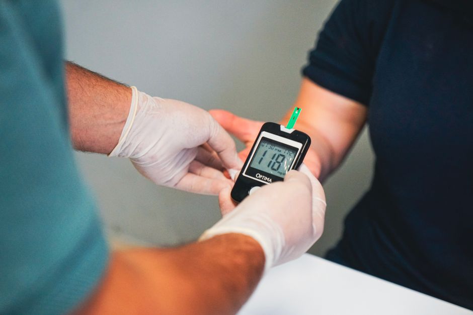
Blood sugar control is the single most important factor for preventing and slowing diabetic retinopathy. Large clinical trials have consistently shown that tighter glucose control reduces the risk of retinopathy onset and progression in people with diabetes.
Blood pressure control reduces stress on retinal vessels. High blood pressure is a well-established risk factor for retinal vein occlusion and hypertensive retinopathy.
Cholesterol management is generally recommended as part of cardiovascular risk reduction, which indirectly supports vascular health throughout the body, including the eye. The direct evidence for cholesterol’s role in retinal vessel disease specifically is less robust than for blood sugar and blood pressure.
Not smoking matters. Smoking damages blood vessels throughout the body and is associated with worse outcomes in several eye diseases.
Treatment depends on the specific condition:
- Diabetic macular edema is commonly treated with anti-VEGF injections into the eye, which reduce leakage and swelling. Laser therapy may also be used in certain cases.
- Proliferative diabetic retinopathy may be treated with panretinal photocoagulation (laser treatment) or anti-VEGF injections, depending on the clinical situation.
- Retinal vein occlusion may be managed with anti-VEGF injections, steroid implants, or laser, depending on the degree of swelling and the presence of abnormal vessel growth.
- Retinal artery occlusion requires emergency evaluation. Treatment options are limited and time-sensitive, and outcomes vary.
These treatments aim to preserve existing vision and slow progression. They do not restore retinal tissue that has already been permanently damaged. This is why early detection through regular eye exams remains the most practical strategy.
What Does the Evidence Actually Show About Retinal Vessel Health?
The link between retinal vessel changes and systemic disease is well established. Research has consistently shown that retinal vessel abnormalities visible on eye exams correlate with cardiovascular risk factors including hypertension and diabetes. Some studies suggest that certain retinal vessel changes may predict cardiovascular events independently of traditional risk factors, though this area continues to be investigated and is not yet part of routine clinical decision-making.
What is not supported by evidence: supplements or lifestyle products marketed specifically for “retinal blood vessel health.” No large human trials have confirmed that any dietary supplement prevents or treats retinal vessel disease. The interventions with demonstrated benefit are management of underlying conditions — blood sugar, blood pressure, cholesterol — and timely treatment of the eye disease itself.
Frequently Asked Questions
Can retinal blood vessel damage be reversed?
Some damage can be stabilized or partially improved with treatment, but retinal tissue that has died cannot be restored. Early treatment gives the best chance of preserving remaining vision.
What causes retinal blood vessels to burst?
Bursting usually results from vessel walls weakened by diabetes, high blood pressure, or abnormal vessel growth. Trauma and certain blood disorders can also cause retinal bleeding.
Can an eye exam detect high blood pressure?
Yes. Retinal vessel changes such as narrowing, crossing abnormalities, and small hemorrhages are visible on a dilated eye exam and can suggest chronic high blood pressure. An eye exam alone cannot diagnose hypertension, but it can prompt further evaluation.
How often should I get my eyes checked if I have diabetes?
Guidelines generally recommend a comprehensive dilated eye exam at least once a year for most people with diabetes, though the right schedule depends on your individual situation. Ask your eye care professional what interval is appropriate for you.
