
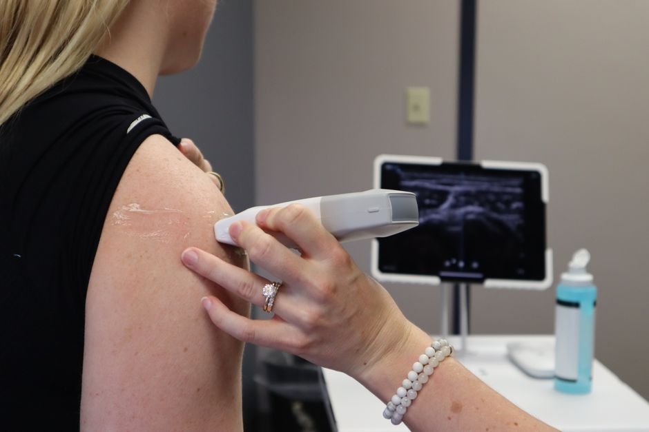
An ultrasound machine sends a short pulse of high-frequency sound waves into the body. When those sound waves hit different tissues, some of them bounce back as echoes. The machine measures how long each echo takes to return and uses that information to build a real-time image on a screen.
How Does an Ultrasound Work From Pulse to Image?
The process starts with a handheld device called a transducer. Inside the transducer are crystals that vibrate when electricity passes through them. Those vibrations produce sound waves at frequencies far above what humans can hear — typically between 2 and 18 megahertz.
The transducer sends out a very short pulse of sound, then immediately switches to listen mode. The pulse travels through the body at a known speed — roughly 1,540 meters per second in soft tissue. When the sound wave encounters a boundary between two different types of tissue, some energy reflects back toward the transducer. The rest continues deeper until it hits another boundary or fades away.
The transducer detects the returning echoes and converts them back into electrical signals. The machine then calculates the distance each echo traveled based on how long it took to return. By processing thousands of these pulses per second and plotting the echo locations, the system creates a two-dimensional image of the internal structures.
What Happens When the Ultrasound Pulse Hits Tissue?
Different tissues reflect sound differently. Bone reflects most of the sound, so it appears bright white on the image — this is called hyperechoic. Fluid, like urine in the bladder or amniotic fluid, reflects very little sound, so it appears black — anechoic. Soft organs like the liver show varying shades of gray depending on their density and structure.
Some sound is absorbed as heat as it passes through tissue, but the amount is very small and well within safety limits for standard diagnostic use. The machine uses a setting called the thermal index to monitor the potential heating effect, especially during long scans or fetal imaging.
The strength of the echo also depends on the angle of the sound beam. If the beam hits a surface at a perpendicular angle, more sound reflects back. If it hits at an angle, more sound scatters or continues deeper. This is why some structures are only visible when the transducer is positioned in a specific way.
How Does the Machine Turn Echoes Into a Picture?
The ultrasound machine uses a process called beamforming to organize the signals. Instead of firing all the crystals at once, the system fires them in a carefully timed sequence. This allows the beam to be focused at a specific depth and steered in a particular direction.
When echoes return, the machine applies a technique called time-gain compensation. Sound waves weaken naturally as they travel deeper, so echoes from deeper tissues are normally weaker. The machine automatically amplifies deeper signals and reduces amplification from shallow signals to produce a uniform brightness across the image.
Each pulse takes a tiny fraction of a second. A typical ultrasound machine sends and receives thousands of pulses to create a single frame. Modern machines can produce 30 to 60 frames per second, which is why the image appears to move in real time.





What Are the Different Types of Ultrasound Imaging?
2D (B-mode) ultrasound is the most common type. It creates a flat, cross-sectional grayscale image of the tissues. This is what you typically see during a pregnancy scan or an abdominal exam.
M-mode ultrasound captures motion along a single line over time. It is often used to measure the movement of heart valves or fetal heart rate because it provides precise timing information.
Doppler ultrasound uses the Doppler effect to measure blood flow. When sound waves hit moving red blood cells, the frequency of the returning echo shifts slightly. The machine calculates the direction and speed of flow from this shift and displays it as color overlays on the 2D image — red typically means flow toward the transducer, blue means flow away.
3D and 4D ultrasound are special types that reconstruct multiple 2D slices into a three-dimensional volume. 4D adds real-time motion. These are not necessary for most medical diagnoses and are sometimes offered for keepsake images, though medical organizations generally advise against non-medical use.
Is Ultrasound Safe? What Are the Risks?
Diagnostic ultrasound has been used for decades with an excellent safety record. It does not use ionizing radiation like X-rays or CT scans. Large population studies have found no convincing evidence of harm from standard diagnostic ultrasound.
There are two theoretical risks the machine monitors. The first is tissue heating from absorbed sound energy. The second is a phenomenon called cavitation, where sound waves cause tiny gas bubbles in tissue to vibrate or collapse. Modern machines display the thermal index (TI) and mechanical index (MI) on screen so the operator can minimize exposure, especially during fetal scans.
However, no large long-term studies have definitively ruled out very subtle effects from prolonged or frequent exposure. For this reason, guidelines recommend using ultrasound only when there is a medical reason and keeping scan times as short as possible. Keepsake ultrasound sessions for non-medical purposes are discouraged by the FDA and other health authorities.
What Can Affect the Quality of an Ultrasound Image?
Several factors can make an ultrasound image harder to interpret. Gas in the bowel blocks sound waves completely, creating dark shadows that obscure deeper structures. This is why patients are sometimes asked to fast before an abdominal ultrasound.
Bone also blocks sound, which is why ultrasound is not useful for examining the brain or spinal cord in adults. In infants, however, the soft spots (fontanelles) provide a window through the skull. Dense scar tissue or surgical hardware can also create artifacts.
The skill of the sonographer matters greatly. Positioning the transducer at the right angle, adjusting gain settings, and knowing how to work around obstacles all affect image clarity. A good sonographer can often get useful images even in difficult situations, while a poor one might miss important findings.
When Is Ultrasound Used vs. Other Imaging Methods?
Ultrasound is often the first imaging test for soft tissues because it is fast, portable, and does not use radiation. Common uses include pregnancy monitoring, gallbladder and kidney stone detection, thyroid and breast mass evaluation, and guiding needle biopsies.
CT scans are better for viewing bone detail, the lungs, and acute bleeding. MRI provides superior detail for soft tissues like the brain, spinal cord, and joints but takes longer and is more expensive. X-rays are best for checking bones and the lungs.
Ultrasound has limitations: it cannot see through bone or gas, and the image quality depends heavily on the operator. For some conditions, ultrasound is the first step and other imaging is used only if the ultrasound result is unclear.
Frequently Asked Questions
Does an ultrasound hurt?
No, an ultrasound is painless. You may feel mild pressure from the transducer on your skin, but there is no pain from the sound waves themselves.
How long does an ultrasound take?
Most diagnostic ultrasound exams take 15 to 45 minutes. The time depends on the body area being scanned and how much detail the doctor needs.
Can ultrasound see all types of cancer?
No. Ultrasound can detect some tumors, especially in soft organs like the liver, kidneys, or thyroid, but it cannot reliably see all cancers. Small tumors, tumors hidden behind bone or gas, and cancers in certain locations may be missed.
Is it safe to have many ultrasounds during pregnancy?
Current evidence shows no harm from standard prenatal ultrasound when used for medical reasons. However, guidelines recommend using it only when necessary for medical care, not for keepsake images or frequent non-medical scans.
