
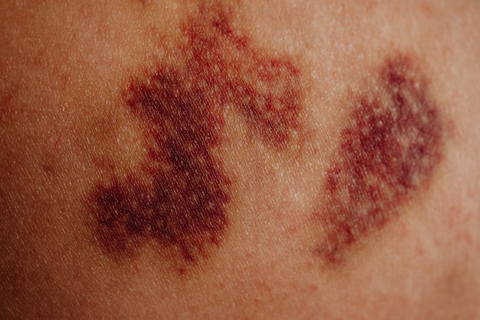
A thrombus forms when the blood clotting system activates inside a living blood vessel and builds a solid mass that stays where it was made. This process involves platelets, clotting proteins, and the vessel wall working together. It is different from a clot that forms in a test tube or a bruise under the skin. Understanding how a thrombus forms helps explain why clots cause heart attacks, strokes, and other serious problems.
How Does A Thrombus Form In A Blood Vessel?
A thrombus forms through a sequence of steps that begin with injury to the vessel lining or abnormal blood flow. The body’s clotting system is designed to seal breaks in blood vessels. When that system activates inside a vessel that has not been cut open, the result is a thrombus.
The process typically follows this order:
- Vessel wall injury or dysfunction. The endothelium is the thin layer of cells lining every blood vessel. When it is damaged or becomes dysfunctional, underlying tissue gets exposed. Platelets stick to that exposed surface.
- Platelet activation and clumping. Once platelets adhere, they change shape and release chemical signals that recruit more platelets to the site. This forms an initial platelet plug.
- Coagulation cascade activation. Clotting proteins in the blood, called coagulation factors, activate in a chain reaction. This cascade converts fibrinogen into fibrin, a mesh-like protein that strengthens the platelet plug.
- Clot stabilization. Fibrin strands weave through the platelet mass, trapping red blood cells and creating a stable thrombus.
This sequence is well established in medical literature. The three conditions that most reliably trigger thrombus formation are known as Virchow’s triad: injury to the vessel wall, abnormal blood flow (stasis or turbulence), and increased tendency of the blood to clot. All three were described by Rudolf Virchow in the 1850s and remain the framework clinicians use today.
What Triggers A Thrombus To Form?
Most thrombi form because one or more parts of Virchow’s triad are present. The trigger is often a combination of factors rather than a single cause.
Vessel wall injury can come from physical damage, high blood pressure, smoking, or the buildup of atherosclerotic plaque. When plaque ruptures, it exposes material that blood is not supposed to contact directly. Platelets respond immediately.
Abnormal blood flow matters because normal flow keeps clotting factors diluted and moving. When blood pools or moves in a chaotic pattern, clotting factors concentrate. This is why atrial fibrillation raises stroke risk. The upper chambers of the heart do not squeeze effectively, so blood stagnates and can form a thrombus. This is also why long periods of immobility, such as after surgery or during extended travel, can lead to clots in the deep veins of the legs.
Hypercoagulability means the blood clots more easily than normal. This can be inherited or acquired. Inherited conditions include factor V Leiden and prothrombin gene mutations. Acquired causes include cancer, pregnancy, certain medications, and inflammatory conditions. Some estimates suggest that inherited clotting disorders are present in a meaningful share of people who develop unprovoked blood clots, though the exact proportion varies by population and study design.
It is worth noting that many people have risk factors and never develop a thrombus. And some people develop thrombi with no identifiable trigger. The relationship between risk factors and clot formation is real but not deterministic.
Arterial vs. Venous Thrombi: What Is The Difference?
Thrombi form in two main types of blood vessels, and the consequences differ substantially.
Arterial thrombi form in arteries, which carry blood away from the heart under high pressure. They are usually triggered by ruptured atherosclerotic plaque. Because arteries feed oxygen to organs, an arterial thrombus can quickly block blood supply. In the heart, this causes a heart attack. In the brain, it causes an ischemic stroke.
Venous thrombi form in veins, where blood pressure is lower and flow is slower. They often start in the deep veins of the legs, a condition called deep vein thrombosis. The main danger is that part of the thrombus can break off and travel to the lungs, causing a pulmonary embolism.
| Feature | Arterial Thrombus | Venous Thrombus |
|---|---|---|
| Typical location | Coronary, cerebral, or peripheral arteries | Deep veins of legs or pelvis |
| Common trigger | Plaque rupture | Stasis, surgery, immobility, hypercoagulability |
| Main complication | Heart attack, stroke | Pulmonary embolism |
| Platelet involvement | High | Lower |
| Fibrin involvement | Present | Dominant |
Arterial thrombi tend to be platelet-rich, which is why antiplatelet drugs like aspirin are used in arterial disease. Venous thrombi are more fibrin-rich, which is why anticoagulants are the main treatment for venous clots. This distinction is one of the more clinically useful things to understand about how thrombi differ.



What Are The Symptoms Of A Thrombus?
Symptoms depend entirely on where the thrombus forms and how much blood flow it blocks.
An arterial thrombus in the heart can cause chest pain, shortness of breath, pain radiating to the arm or jaw, and sweating. In the brain, it can cause sudden weakness on one side, difficulty speaking, vision changes, or loss of coordination.
A venous thrombus in the leg may cause swelling, pain, warmth, and redness in the affected limb. But many deep vein thrombi cause no symptoms at all. This is a critical point. A clot can be present and growing without any warning signs.
A pulmonary embolism can cause sudden shortness of breath, chest pain that worsens with breathing, rapid heart rate, and coughing up blood. This is a medical emergency.
If you experience symptoms that could indicate a stroke or heart attack, call emergency services immediately. Time matters for treatment.
How Is A Thrombus Diagnosed And Treated?
Diagnosis depends on the location and clinical suspicion. For deep vein thrombosis, ultrasound is the standard first test. For pulmonary embolism, CT angiography is commonly used. For arterial thrombi, imaging may include angiography or other vascular studies. Blood tests such as D-dimer can help rule out clots in low-risk situations, but they are not definitive on their own.
Treatment aims to stop the thrombus from growing, prevent it from breaking off, and reduce the risk of new clots. The approach varies by type and location.
- Anticoagulants slow or stop the clotting process. They are the main treatment for venous thrombi. Common options include heparin, warfarin, and direct oral anticoagulants.
- Antiplatelet drugs reduce platelet activity and are used mainly for arterial thrombi. Aspirin is the most well-known example.
- Thrombolytic drugs dissolve existing clots and are used in emergencies such as major strokes or large pulmonary emboli. They carry a higher bleeding risk.
- Procedures such as thrombectomy may be used to physically remove a clot in certain situations.
Treatment decisions are made by clinicians based on the specific situation. The evidence supporting anticoagulation for venous thromboembolism is strong and consistent across many trials. The choice of which anticoagulant, and for how long, depends on factors including the cause of the clot, bleeding risk, and patient preference.
Can A Thrombus Be Prevented?
Prevention focuses on reducing the conditions that allow thrombi to form. Not all clots are preventable, but many risk factors can be addressed.
For people at risk of venous thromboembolism, movement is important. After surgery or during long periods of sitting, getting up and moving the legs helps keep blood flowing. Compression stockings are sometimes used. In hospital settings, preventive anticoagulation is standard for many surgical patients and those with limited mobility.
For arterial disease, managing blood pressure, cholesterol, and blood sugar matters. Not smoking reduces vessel injury. Regular physical activity supports healthy circulation. These measures are supported by strong evidence for reducing cardiovascular events, though they do not eliminate clot risk entirely.
If you have a personal or family history of blood clots, or a known clotting disorder, talk to your doctor about your risk. Some people benefit from long-term anticoagulation, but this decision requires weighing benefits against bleeding risk. There is no single answer that applies to everyone.
What Is The Difference Between A Thrombus And An Embolus?
A thrombus stays where it forms. An embolus is a clot or other material that travels through the bloodstream and blocks a vessel somewhere else. A thrombus can become an embolus if part of it breaks loose.
This distinction matters because the danger of a venous thrombus in the leg is not usually the clot itself. It is that a piece may travel to the lungs. Similarly, a thrombus in the heart can embolize to the brain and cause a stroke.
The word “embolism” refers to the process of blockage by traveling material. The material itself is called an embolus. A thrombus that has embolized is sometimes called a thromboembolism.
Frequently Asked Questions
How long does it take for a thrombus to form?
A thrombus can begin forming within seconds to minutes after vessel injury or trigger. The full clot may develop over hours to days depending on the conditions.
Can a thrombus go away on its own?
Small thrombi can sometimes dissolve naturally through the body’s fibrinolytic system. Larger or more dangerous clots usually require medical treatment.
What is the most common cause of a thrombus?
The most common underlying causes are vessel wall injury, abnormal blood flow, and hypercoagulability. These three factors make up Virchow’s triad.
Can you feel a thrombus forming?
Not always. Many thrombi cause no symptoms until they block blood flow or break off. Some cause pain, swelling, or warmth in the affected area.
