
Finding out you have a lung lesion can be unsettling, but it does not automatically mean you have cancer. A lung lesion is simply an abnormal spot or area of tissue in the lung, and the vast majority of these findings are benign, or non-cancerous. The seriousness of a lung lesion depends entirely on its size, shape, location, and your personal risk factors like age and smoking history, which is why a structured evaluation by a doctor is the only way to determine what it is and whether treatment is necessary.
What Exactly Is a Lung Lesion?
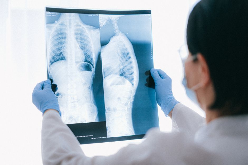
A lung lesion is a broad term for any area of abnormal tissue within the lungs. It can look like a nodule, a mass, or an area of inflammation on an imaging scan. When doctors use the term “lesion,” they are describing what they see on a chest X-ray or CT scan before they know the cause.
These spots are often found by accident. You might have a CT scan for an unrelated reason, like a heart check or a car accident, and the radiologist notices a spot on your lung. This is incredibly common. As CT scans have become more detailed, doctors find more of these spots than ever before, many of which are harmless.
The key distinction doctors make is between a nodule and a mass. A nodule is typically defined as a spot smaller than 3 centimeters (about 1.2 inches). A mass is larger than 3 centimeters. Size matters because larger lesions carry a higher risk of being cancerous, though size alone does not tell the whole story.
What Causes Lung Lesions?
There are many causes of lung lesions, and most are not cancer. The cause often relates to past infections, inflammation, or benign growths.
Common benign causes include:
- Granulomas: Small areas of inflammation caused by past infections like tuberculosis or fungal infections. These are very common and usually harmless.
- Hamartomas: Benign tumors made of normal lung tissue that grew in a disorganized way. They are the most common type of benign lung tumor.
- Scar tissue: Areas of fibrosis from a prior pneumonia, injury, or surgery.
- Infections: Active infections like bacterial pneumonia or bronchitis can create temporary opacities or lesions on imaging.
- Pulmonary nodules: Many small, non-cancerous nodules can develop from exposure to dust, fumes, or autoimmune conditions.
Malignant causes include primary lung cancer, which starts in the lung, and metastatic disease, which means cancer from another part of the body has spread to the lungs. Your doctor will consider your full medical history when weighing these possibilities.
Are Lung Lesions Serious, Causes, Evaluation, and Treatment: When to Worry
The honest answer is that seriousness is determined by evaluation, not by the word “lesion” itself. Certain features on a CT scan make a lesion more concerning for cancer.
A lesion is more suspicious if it is large, has irregular or spiculated borders (meaning it looks like it has spikes radiating out), or if it grows on a follow-up scan. Calcified lesions, which contain calcium deposits, are usually benign. Lesions that remain the same size for two years on serial scans are also almost always benign.
Your personal risk factors are just as important as the scan image. A history of heavy smoking, older age, a personal history of cancer, or a family history of lung cancer all increase the risk that a lesion is malignant. Conversely, a young person with no smoking history who has a small, smooth nodule has a very low risk of cancer.
This is why a single scan is rarely enough to make a final judgment. The standard of care often involves serial imaging to check for growth or stability.




How Are Lung Lesions Evaluated?
Evaluation starts with a careful review of the imaging. Your doctor will look at the exact characteristics of the lesion and compare it to any old scans you might have. If you have a previous scan from years ago, that is the single most valuable tool for determining if the lesion is new or stable.
If the lesion looks low-risk, the typical approach is active surveillance. This means you will get another CT scan in a set timeframe, often 3, 6, or 12 months later, depending on the lesion’s size and your risk profile. The goal is to see if it changes over time. Benign lesions stay the same or shrink. Cancerous lesions usually grow.
If the lesion looks high-risk, or if it grows on a follow-up scan, your doctor will likely recommend a biopsy. There are several ways to biopsy a lung lesion:
- Bronchoscopy: A thin, flexible tube is passed through your mouth or nose into your airways to reach the lesion.
- CT-guided needle biopsy: A needle is inserted through your chest wall into the lesion using CT imaging for guidance.
- Surgical biopsy: Sometimes a small sample is taken during a minimally invasive procedure called video-assisted thoracoscopic surgery.
In some cases, a PET scan may be used. This scan measures metabolic activity. Cancerous cells often use more sugar than normal cells, so they light up on a PET scan. However, inflammation can also cause PET scans to light up, so this test is not definitive on its own.
Treatment Options for Lung Lesions
Treatment depends entirely on the diagnosis. There is no blanket treatment for a “lung lesion” because the term covers so many different conditions.
If the lesion is benign, no treatment is needed. You may simply need periodic follow-up scans to confirm it remains stable, and then you will be discharged from monitoring. Granulomas, hamartomas, and scar tissue do not require surgery or medication.
If the lesion is an active infection, you will be treated with the appropriate antibiotics or antifungal medications. Once the infection clears, the lesion may disappear entirely or leave behind harmless scar tissue.
If the lesion is malignant, treatment depends on the type and stage of the cancer. Options may include surgical removal of the lesion and surrounding tissue, radiation therapy, chemotherapy, targeted therapy, or immunotherapy. The specific plan is customized by an oncology team based on biopsy results and staging scans.
What Happens During Follow-Up Monitoring?
Follow-up monitoring is not optional when it comes to lung lesions. It is a critical part of the evaluation process because it provides the data needed to determine if a lesion is growing.
Missing a scheduled follow-up scan is the most common reason a benign lesion turns into a delayed cancer diagnosis. If a lesion is going to grow, it usually does so slowly, over months or years. Serial CT scans catch that growth early, when treatment is most effective.
The specific follow-up interval is based on published guidelines from radiology and pulmonology societies. These guidelines consider the size of the nodule and your individual risk factors. A small nodule in a low-risk person might only need one follow-up scan at 12 months. A larger nodule in a high-risk person might need scans every 3 to 6 months for the first two years.
Your doctor will give you a clear schedule. It is important to keep those appointments even if you feel perfectly fine. Lung lesions rarely cause symptoms when they are small, so you cannot rely on how you feel to know if something has changed.
Can Lung Lesions Cause Symptoms?
Most lung lesions cause no symptoms at all. They are discovered incidentally on imaging done for other reasons. This is why routine screening for high-risk individuals is so important—it catches lesions before they cause problems.
When lesions do cause symptoms, it is usually because they are large, located near an airway, or have spread. Possible symptoms include a persistent cough, coughing up blood, chest pain, shortness of breath, or recurrent respiratory infections. Unexplained weight loss and fatigue can also occur with malignant lesions.
If you experience any of these symptoms, especially if you are a current or former smoker, you should see a doctor promptly. However, these symptoms are not specific to lung lesions and can be caused by many other conditions.
Frequently Asked Questions
Can a lung lesion be benign?
Yes, most lung lesions are benign. Granulomas, scar tissue, and hamartomas are common non-cancerous causes found on imaging scans.
How fast do lung lesions grow?
Growth rates vary widely depending on the cause, but cancerous lesions typically double in size over several months to a couple of years. This is why follow-up scans are scheduled months apart to detect meaningful changes.
Is a biopsy always needed for a lung lesion?
No, a biopsy is only needed if the lesion looks suspicious or grows on follow-up scans. Many low-risk lesions are managed with regular imaging alone.
What size lung lesion is considered dangerous?
Lesions larger than 3 centimeters, called masses, carry a higher cancer risk than smaller nodules. However, size alone does not determine danger, and even small lesions can be malignant in high-risk individuals.
