
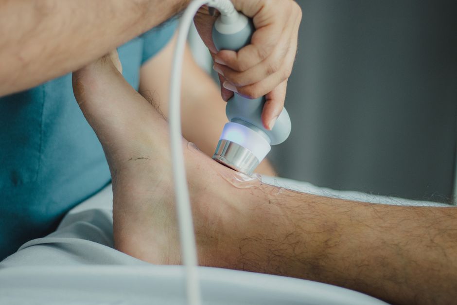
A retroperitoneal ultrasound is done to look at organs located deep in the back of the abdomen, behind the membrane that lines the abdominal cavity. Doctors order this scan to check for problems in the kidneys, pancreas, aorta, and other structures that sit in this space. It is a painless, radiation-free imaging test that helps diagnose pain, detect masses, or monitor known conditions.
What Exactly Is the Retroperitoneum?
The retroperitoneum is the space in the back of your belly, behind the peritoneum. The peritoneum is a thin membrane that wraps around most of your abdominal organs. Organs in the retroperitoneum sit behind that membrane, pressed against your back muscles and spine.
This space contains several important structures. The kidneys sit here, along with the ureters, the tubes that carry urine from the kidneys to the bladder. The pancreas lies mostly in the retroperitoneum. The abdominal aorta, the largest artery in the body, runs through this space. The adrenal glands, which sit on top of each kidney, are also retroperitoneal.
Because these organs are deep inside the body, they cannot be felt during a physical exam. An ultrasound gives doctors a way to see them without surgery or radiation exposure.
Why Is A Retroperitoneal Ultrasound Done? Why It Happens
A doctor orders a retroperitoneal ultrasound when symptoms point to a problem in one of these deep organs. The most common reasons include flank pain, blood in the urine, suspected kidney stones, or an abnormal blood test that suggests kidney trouble.
The test is also used to look for masses. If a CT scan or MRI found something suspicious in the abdomen, an ultrasound can provide more detail about whether it is solid or filled with fluid. This helps doctors decide if a biopsy is needed.
For people with a family history of abdominal aortic aneurysm, ultrasound is the standard screening tool. The aorta can widen silently without causing symptoms. An ultrasound measures its width and can catch an aneurysm before it becomes dangerous.
Pancreatic problems are another reason for this scan. If someone has persistent upper abdominal pain that radiates to the back, or unexplained weight loss, the doctor may order a retroperitoneal ultrasound to look at the pancreas. Ultrasound can detect pancreatic masses or fluid collections, though it has limits in fully seeing this organ.
Doctors also use this test to guide procedures. If a fluid collection needs to be drained or a biopsy needs to be taken from a kidney, ultrasound provides real-time images to guide the needle safely.
How the Test Works and What to Expect
A retroperitoneal ultrasound uses sound waves to create images. A handheld device called a transducer sends sound waves into your body. These waves bounce off organs and return to the transducer, which converts them into a picture on a screen.
You lie on your back for most of the exam. The sonographer applies a water-based gel to your abdomen to help the sound waves travel smoothly. The transducer is pressed against your skin and moved around to capture images from different angles.
You may be asked to roll onto your side. This position can improve the view of the kidneys. You might also need to hold your breath briefly during some images. Breath-holding reduces movement and gives a clearer picture.
The test typically takes 30 to 45 minutes. There is no radiation involved, no needles, and no recovery time. You can drive yourself home and return to normal activities immediately after.
Preparation is usually minimal. Some doctors ask you to fast for six to eight hours before the test. An empty stomach reduces gas in the bowel, which can block sound waves and obscure the organs behind it. You may also be asked to drink water before the exam to fill your bladder, which helps push bowel loops out of the way.





What the Ultrasound Can and Cannot Detect
Ultrasound is excellent at telling whether a mass is solid or cystic. A simple cyst filled with clear fluid is often benign and needs no further testing. A solid mass may require a biopsy to determine if it is cancerous.
Kidney stones appear on ultrasound, though small stones can be missed. CT scans are more sensitive for detecting tiny stones. If ultrasound shows no stone but symptoms persist, the doctor may order a CT scan for a more detailed look.
Ultrasound measures blood flow using a technique called Doppler. This shows how well blood is moving through the kidneys and the aorta. Narrowed blood vessels or blockages can be detected this way.
Hydronephrosis, a condition where urine backs up and swells the kidney, is clearly visible on ultrasound. The dilated collecting system appears as dark spaces within the kidney. This finding helps doctors determine if a stone or tumor is blocking urine flow.
The pancreas is harder to evaluate with ultrasound. Gas in the stomach and intestines often blocks the view. If the pancreas cannot be seen clearly, the doctor may order a CT scan or MRI instead. Ultrasound remains useful for detecting advanced pancreatic masses or fluid collections.
Ultrasound cannot reliably detect small tumors in the retroperitoneum. It also cannot determine whether a mass is cancerous with certainty. That determination requires a biopsy and pathology review.
Conditions That Prompt This Scan
Several specific conditions lead doctors to order a retroperitoneal ultrasound. Renal cell carcinoma, a type of kidney cancer, may be found when investigating blood in the urine or flank pain. Ultrasound can show the tumor and help stage the disease.
Polycystic kidney disease, an inherited condition where fluid-filled cysts grow in the kidneys, is monitored with periodic ultrasounds. The scans track cyst growth and kidney size over time.
An abdominal aortic aneurysm is a bulging, weakened area in the aorta. Screening ultrasound is recommended for men aged 65 to 75 who have ever smoked. The test measures the aorta diameter. A normal aorta is about 2 centimeters wide. An aneurysm is diagnosed when the diameter reaches 3 centimeters or more.
Retroperitoneal fibrosis is a rare condition where fibrous tissue grows in the retroperitoneum, often wrapping around the ureters and blocking urine flow. Ultrasound can show the resulting hydronephrosis and help guide treatment.
Lymph nodes in the retroperitoneum can enlarge due to lymphoma or metastatic cancer. Ultrasound may detect these enlarged nodes, though CT and PET scans are more comprehensive for staging cancer.
Limitations and Follow-Up Testing
Ultrasound has real limitations that patients should understand. The images depend heavily on the skill of the sonographer and the quality of the equipment. Body habitus matters too. Excess abdominal fat or gas can degrade image quality significantly.
If the ultrasound is inconclusive, your doctor will likely order a CT scan or MRI. These tests provide more detailed images of the retroperitoneum. CT scans use radiation, while MRI uses magnetic fields. Both can see through gas and fat more effectively than ultrasound.
A normal ultrasound does not always rule out disease. Small masses, subtle abnormalities, or early-stage conditions may not appear on the scan. If your symptoms continue after a normal ultrasound, tell your doctor. Further testing may be necessary.
An abnormal ultrasound finding does not automatically mean cancer or serious disease. Many findings require additional testing before a diagnosis is made. Your doctor will explain what the results mean and what steps come next.
Risks and Safety of the Procedure
Diagnostic ultrasound has no known harmful effects when used appropriately. It does not use ionizing radiation like X-rays or CT scans do. The sound waves are at a frequency too high for human hearing, and the energy levels used for imaging are low.
Ultrasound is safe during pregnancy, which makes it valuable for evaluating pregnant patients with kidney problems. It is also safe for children and for people with pacemakers or metal implants that would prevent an MRI.
The only discomfort is mild pressure from the transducer on your abdomen. If you have abdominal tenderness, tell the sonographer. They can adjust their technique to minimize discomfort.
There are no known cumulative risks from repeated ultrasounds. This makes it a good choice for monitoring chronic conditions that require regular imaging over many years.
Frequently Asked Questions
How long does a retroperitoneal ultrasound take?
The test typically takes 30 to 45 minutes. You can return to normal activities immediately afterward.
Do I need to fast before a retroperitoneal ultrasound?
Many doctors recommend fasting for six to eight hours before the exam. An empty stomach reduces bowel gas that can block the ultrasound images.
Is a retroperitoneal ultrasound painful?
No, the test is generally painless. You may feel mild pressure where the transducer presses against your abdomen, but this should not cause significant discomfort.
Can a retroperitoneal ultrasound detect cancer?
Ultrasound can detect masses that may be cancerous, but it cannot confirm a cancer diagnosis. A biopsy is required to determine if a mass is malignant.
What is the difference between an abdominal ultrasound and a retroperitoneal ultrasound?
An abdominal ultrasound examines organs in the front of the belly, like the liver and gallbladder. A retroperitoneal ultrasound focuses on the deeper organs behind the peritoneum, including the kidneys, pancreas, and aorta.
