
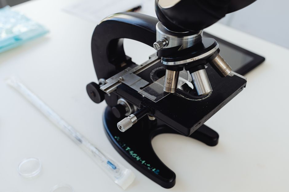
What Makes a Dissecting Microscope Different From a Compound Microscope?
The most important difference is depth perception. A dissecting microscope uses two separate optical paths—one for each eye—that view the specimen from slightly different angles. Your brain combines these images into a single three-dimensional view. A compound microscope uses one optical path, so the image appears flat.
There are practical differences too. Compound microscopes are designed for thin, transparent samples mounted on glass slides. Light shines up through the specimen from below. Dissecting microscopes use reflected light from above or a combination of top and bottom lighting. This means you can look at an object that is solid, thick, or opaque.
Magnification is another major difference. Compound microscopes typically magnify from 40x up to 1000x or more. Dissecting microscopes usually range from 5x to 50x, though some models go higher. That lower magnification is not a weakness—it is what allows you to see the whole object in context rather than just a tiny slice of it.
Working distance is the space between the lens and the specimen. Dissecting microscopes have a long working distance, often several inches. This gives you room to use tools like forceps, probes, and scalpels while you observe. You cannot do that under a compound microscope because the lens sits too close to the slide.
When Would You Use a Dissecting Microscope in Biology and Education?
Biology classrooms are where most people first encounter these instruments. They are ideal for teaching anatomy because students can dissect a specimen and see exactly where they are cutting.
A typical classroom exercise involves examining a flower. Under a dissecting microscope, you can identify the petals, sepals, stamens, and pistil without destroying the flower. You can pull the parts apart and look at their textures and structures. A compound microscope would require you to make thin cross-sections, which is a different skill entirely.
Insects are another common subject. You can examine a butterfly wing and see the individual scales that give it color. You can look at an ant’s body segments, legs, and antennae in detail. Because the insect is intact and three-dimensional, a dissecting microscope is the only practical way to view it.
Dissection of preserved specimens—like frogs, fetal pigs, or earthworms—is almost always done under a dissecting microscope. The long working distance allows students to manipulate the specimen with scissors and probes while seeing their work magnified. This is not just a classroom convenience. It teaches careful technique that carries into professional lab work.
How Are Dissecting Microscopes Used in Electronics and Manufacturing?
Electronics repair and circuit board inspection rely heavily on dissecting microscopes. Modern circuit boards have components that are tiny but still visible to the naked eye. Solder joints, hairline cracks, and damaged traces are another story. A dissecting microscope reveals these details clearly.
Technicians use them for soldering work. The three-dimensional view helps judge depth when placing components or checking whether a solder joint is properly formed. The working distance allows room for a soldering iron. This is common practice in phone repair shops and manufacturing quality control.
Jewelry makers and watch repairers use dissecting microscopes for the same reasons. Setting a small stone or repairing a watch movement requires precise hand-eye coordination. The stereo view lets them see depth that a magnifying lamp cannot provide.
Quality control inspectors in manufacturing use these microscopes to check for surface defects. This includes things like cracks in metal parts, imperfections in plastic molding, or contamination on medical devices. The ability to inspect an object without preparing or destroying it makes the dissecting microscope valuable across industries.
-
 Garlic Benefits for Men: What the Science Actually Shows.
 -
Natural Remedies for Constipation: 10 Science-Backed Options
-
How to Loosen a Bowel Blockage at Home. Easy Tips To Follow
-
Can Intermittent Fasting Cause Constipation? Benefits, Pros & Cons
-
Pulmo Balance Reviews 2026: Legit Lung Support Or Not?





What Can You Examine at Home With a Dissecting Microscope?
Hobbyists and curious families use dissecting microscopes for a wide range of projects. The low magnification and reflected light make them forgiving instruments for beginners. You do not need to prepare slides or stain specimens.
Rocks and minerals are excellent subjects. You can see crystal structures, grain boundaries, and tiny fossils embedded in stone. A dissecting microscope reveals details that a hand lens cannot show.
Coins and stamps show their condition clearly under this type of microscope. Collectors can examine mint marks, wear patterns, and printing details without risking damage to the item. The reflected light brings out surface texture in a way that flat scanning cannot.
Textiles and paper reveal their structure at this magnification. You can see individual fibers in fabric, the weave pattern, and signs of wear or damage. Paper shows its grain and any surface treatments. This has practical uses for conservators and hobbyists alike.
Food science is another home application. Examining the surface of a strawberry or a leaf of lettuce under a dissecting microscope can show you the tiny structures that make them up. You can see the pores on a leaf, called stomata, though you may need higher magnification to see them clearly. The tiny hairs on plant stems, called trichomes, are easy to spot.
What Are the Limitations of a Dissecting Microscope?
A dissecting microscope cannot show you cells. The maximum useful magnification is generally around 50x to 100x. Individual animal cells are typically 10 to 30 micrometers across. You need at least 400x magnification to see them clearly, which is beyond the range of most dissecting microscopes.
You also cannot see through opaque objects. The reflected light shows surface detail only. If you need to see internal structures of a solid object, you must cut it open or use imaging technology like X-rays or MRI.
Depth of field is shallow at higher magnifications. Even though you get a three-dimensional view, only a thin plane of the specimen is in focus at once. You will need to adjust the focus frequently as you move across a curved or uneven surface.
For very small objects, a dissecting microscope is not enough. A single-celled organism like a paramecium is invisible at 20x magnification. Viewing bacteria requires a compound microscope with an oil immersion lens at 1000x. The two instruments do not compete with each other—they serve different purposes.
How Do You Choose the Right Dissecting Microscope?
Consider what you will examine most often. This determines the magnification range you need. For general biology and dissection, a zoom range of 10x to 40x is practical. For electronics work, many technicians prefer a range that goes up to 50x or higher.
Lighting matters more than most beginners expect. Many dissecting microscopes have lights built into the stand, shining from above. Some also have transmitted light from below for examining translucent specimens. If you work with opaque objects like circuit boards or rocks, top lighting is essential. If you examine thin specimens like leaves or insect wings, you may want both options.
Working distance is worth checking before purchase. If you plan to do dissection or soldering, you need enough space to move your tools. Some microscope bodies have a shorter working distance at higher magnification, which can make detailed work difficult.
Eyepieces and viewing comfort are personal but important. Look for a microscope with adjustable eyepieces if you wear glasses. Some models have a diopter adjustment on one eyepiece to compensate for differences between your eyes. A binocular head—one with two eyepieces—is standard and provides the three-dimensional view that makes these instruments useful.
Frequently Asked Questions
Can a dissecting microscope see cells?
No, a dissecting microscope cannot see individual cells clearly. Its magnification is too low, and most cells are transparent and require transmitted light and staining to be visible.
What is the difference between a stereo microscope and a dissecting microscope?
They are the same instrument. The terms are used interchangeably because the two eyepieces provide a three-dimensional, or stereo, view that is ideal for dissection work.
Do I need to prepare slides for a dissecting microscope?
No, you do not need slides for most dissecting microscope work. You place the whole object directly on the stage and view it with reflected light from above.
What magnification is best for a beginner dissecting microscope?
A zoom range of about 10x to 40x is the most practical for general use. This covers insects, plants, coins, and basic dissection without the shallow depth of field found at higher magnifications.
