
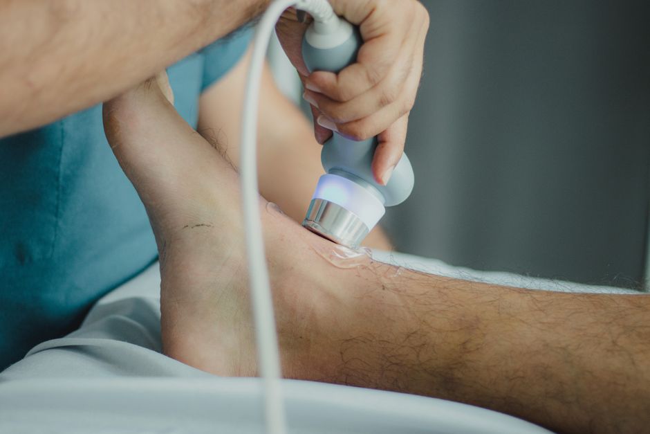
Ultrasound elastography is a medical imaging technique that measures the stiffness of body tissues. It works by sending sound waves into the body and tracking how tissues move in response to a gentle push. This information helps doctors tell the difference between healthy and diseased tissue, such as scarred liver or cancerous tumors.
What Is Ultrasound Elastography And How Does It Work?
Ultrasound elastography is a specialized form of ultrasound that adds information about tissue stiffness to the standard images. Traditional ultrasound shows the shape and structure of organs and blood flow. Elastography adds a color map that displays stiffness, which can reveal disease.
The technique works through two main methods. The first is strain elastography. The operator applies gentle pressure with the ultrasound probe, and the system measures how much the tissue deforms. Softer tissue compresses more; stiffer tissue compresses less. The second method is shear wave elastography. This uses a focused sound pulse to create a tiny wave that travels sideways through the tissue. The system measures the speed of that wave. Stiffer tissue transmits the wave faster.
Both methods produce a numerical value or a color overlay on the screen. Blue often indicates softer tissue, red stiffer. The results can be interpreted immediately by a trained radiologist or sonographer.
Why Is Ultrasound Elastography Used?
Doctors use elastography to evaluate diseases that change the stiffness of organs. The most common use is for the liver. Chronic liver diseases like hepatitis or fatty liver can cause scarring, known as fibrosis. As fibrosis worsens, the liver becomes stiffer. Elastography can measure this stiffness and help stage the disease without needing a biopsy.
Clinicians also use elastography for breast lesions. It helps distinguish benign lumps from suspicious ones. A soft, easily compressible lump is more likely benign. A stiff, hard lump raises concern for cancer. It is not a replacement for biopsy, but it provides additional information during an ultrasound exam.
Other uses include evaluating thyroid nodules, prostate lesions, lymph nodes, and musculoskeletal injuries. Research continues to expand the applications.
What Conditions Can Ultrasound Elastography Help Diagnose?
Liver fibrosis and cirrhosis are the most established uses. Studies consistently show that elastography can detect significant fibrosis and cirrhosis with high accuracy. It reduces the need for liver biopsies in many patients. Some clinical guidelines recommend elastography as the first-line test for liver fibrosis in certain populations.
Breast lesions are another common application. Elastography can increase the specificity of breast ultrasound. That means it helps rule out cancer more confidently when the standard ultrasound looks suspicious. However, suspicious findings still need a biopsy.
Thyroid nodules can be assessed with elastography. Stiffer nodules are more likely to be cancerous. The evidence here is good but not as strong as for liver. Most guidelines use elastography as one factor among many when deciding if a nodule needs a fine-needle aspiration.
Other areas include evaluating prostate cancer, cervical stiffness during pregnancy, and tendon injuries. The evidence in these areas is still emerging. Not all are standard clinical practice.





How Does Ultrasound Elastography Differ From a Regular Ultrasound?
Regular ultrasound uses sound waves to create images of the inside of your body. It shows the size, shape, and texture of organs and can detect cysts, tumors, or stones. It also measures blood flow with Doppler technology.
Elastography adds a completely different type of information: tissue stiffness. A regular ultrasound cannot measure stiffness. Two tissues may look identical on a standard ultrasound but have very different stiffness. For example, a benign cyst and a solid cancer may both appear as a dark spot. On elastography, the cyst is soft and compressible, while the cancer is stiff. This additional data helps doctors make better decisions.
The procedure itself is similar for the patient. You lie on a table, gel is applied to the skin, and a probe is moved over the area. Elastography adds just a few minutes to a standard ultrasound exam.
Is Ultrasound Elastography Safe?
Yes, ultrasound elastography is considered very safe. It uses the same sound wave technology as regular ultrasound, which has no known harmful effects when used properly. There is no radiation involved.
The gentle push used in strain elastography is just pressure from the probe. Shear wave elastography uses a focused pulse that is stronger than standard ultrasound but still within approved safety limits. No complications have been reported from the procedure itself.
That said, the technique requires training and skill. Results depend on the operator and the patient’s body habitus. In some people, especially those with a large amount of abdominal fat, the stiffness measurements may be less reliable. Your doctor will interpret the results in the context of your full medical picture.
What Should I Expect During an Ultrasound Elastography Procedure?
The exam is very similar to a standard ultrasound. You will be asked to change into a gown and lie on an exam table. A gel is applied to the skin over the area being studied. The sonographer moves a handheld probe over the skin. You may feel light pressure but it should not be painful.
During strain elastography, the sonographer may gently push the probe in and out several times. This can feel like a slight bounce. During shear wave elastography, you may feel a brief deep vibration or tap. It is not uncomfortable. The entire exam usually takes 15 to 30 minutes for a single organ.
After the exam, the gel is wiped off, and you can go back to normal activities. A radiologist or your doctor will review the images and stiffness measurements. Results are typically available within a few days.
Frequently Asked Questions
Does ultrasound elastography hurt?
No, it does not hurt. You may feel slight pressure from the probe or a gentle tapping sensation during shear wave measurements. Most people find it comfortable.
How accurate is elastography for liver disease?
Elastography is quite accurate for detecting significant liver fibrosis and cirrhosis. It is less reliable for very early stages of fibrosis. Some factors like obesity or fluid in the abdomen can affect accuracy.
Can elastography replace a biopsy?
In many cases of liver disease, yes, elastography can replace a biopsy for staging fibrosis. For breast or thyroid lesions, it provides helpful information but does not replace a biopsy when cancer is suspected.
Is elastography covered by insurance?
Coverage varies by insurance plan and medical indication. Liver elastography is often covered for chronic liver disease. Other uses may require preauthorization. Check with your insurance provider.
