
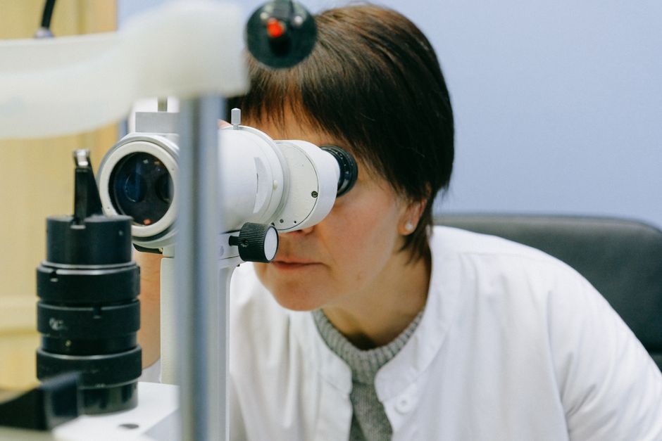
Retinal tears most commonly occur when the vitreous gel inside the eye pulls away from the retina as a normal part of aging. This process, called posterior vitreous detachment (PVD), happens gradually in most people by their 70s or 80s. When the vitreous separates from the retina, it can tug hard enough to tear the retinal tissue, creating a retinal tear.
What Is the Most Common Cause of Retinal Tear?
The most common cause of a retinal tear is posterior vitreous detachment (PVD). As you age, the vitreous gel that fills the eye naturally shrinks and liquefies. Over time, it separates from the retina, the light-sensitive layer at the back of the eye. In most people this separation happens without problems. But in some cases, the vitreous remains firmly attached to certain spots on the retina. When it pulls away, it can rip a small hole or tear in the retina.
PVD is a normal age-related change. It typically begins after age 50 and becomes more common after age 60. By age 80, most people have some degree of PVD. Not everyone who has PVD develops a retinal tear, but this detachment is the single most common trigger for a tear.
What Happens During Posterior Vitreous Detachment?
To understand why PVD causes tears, it helps to know the eye’s anatomy. The vitreous is a clear, jelly-like substance that fills the space between the lens and the retina. It contains millions of fine fibers that attach to the retina. With age, the vitreous shrinks and the fibers can break. Normally, the vitreous slips away from the retina smoothly.
When the fibers are unusually strong or the vitreous is especially sticky, it pulls the retina as it separates. That stretching can create a full-thickness break, which is a retinal tear. The tear allows fluid from the vitreous cavity to seep underneath the retina. If enough fluid collects, the retina can begin to lift off the back of the eye, leading to a retinal detachment—a vision-threatening emergency.
Most retinal tears from PVD occur in the peripheral (side) part of the retina. They rarely happen in the central area (the macula) unless other factors are involved.
Are There Other Causes of Retinal Tears?
While PVD is the most common cause, other factors can also lead to retinal tears. Eye injuries from blunt trauma, such as a punch or a fall, can directly tear the retina. Diabetic retinopathy can cause scar tissue that pulls on the retina, sometimes creating a tear. High levels of nearsightedness (myopia) increase the risk because the eye is longer and the retina is thinner and more fragile.
Retinal tears can also occur after eye surgery, particularly cataract surgery, because the removal of the lens can shift the vitreous slightly and trigger a PVD. Inflammation inside the eye (uveitis) and certain genetic conditions that affect the retina can also cause tears, but these are far less common than age-related PVD.
No single cause besides PVD accounts for the majority of retinal tears. Most cases seen by eye doctors are linked to aging and vitreous changes.





What Are the Symptoms of a Retinal Tear?
Recognizing symptoms early is critical. A retinal tear often causes sudden onset of floaters—small dark specks or cobweb-like shapes that drift across your vision. You may also see flashes of light, especially in the corner of your eye. These flashes occur when the vitreous tugs on the retina, stimulating it.
Another symptom is a shadow or curtain that appears in part of your visual field. This suggests that fluid is collecting under the retina and a detachment may be starting. If you notice any of these signs, you need an urgent eye exam. A retinal tear can be treated effectively if caught early. Waiting can allow it to progress to a retinal detachment, which may require surgery and can cause permanent vision loss.
It is possible to have a retinal tear without symptoms, especially if the tear is small and located far from the center of vision. This is why routine dilated eye exams are important, particularly if you have risk factors like high nearsightedness or a family history of retinal problems.
How Is a Retinal Tear Treated?
Treatment for a retinal tear aims to seal the tear and prevent fluid from getting under the retina. The two most common procedures are laser photocoagulation and cryopexy. In laser treatment, a doctor uses a laser to create small burns around the tear. This creates scar tissue that seals the retina to the underlying tissue. In cryopexy, a freezing probe is applied to the outside of the eye to freeze the area around the tear and form a scar.
Both treatments are performed in an office setting and are usually successful in preventing progression to retinal detachment. The choice between them depends on the location and size of the tear, as well as the doctor’s preference. After treatment, you will need follow-up exams to confirm the seal holds. In some cases, a second treatment may be needed if the tear persists.
If a retinal detachment has already started, surgery such as a vitrectomy or scleral buckle may be required. The earlier the tear is treated, the simpler the procedure and the better the visual outcome.
Can You Prevent a Retinal Tear?
There is no proven way to prevent posterior vitreous detachment because it is a natural aging process. However, you can reduce your risk of a tear by protecting your eyes from trauma. Wear safety goggles during sports or activities that could cause eye injury. If you have diabetes, keeping your blood sugar under control can help prevent diabetic retinopathy, which can lead to retinal tears.
Regular eye exams are important, especially if you are nearsighted, have had cataract surgery, or have a family history of retinal problems. A dilated exam allows an eye doctor to see the retina clearly and may detect a tear before it causes symptoms. While you cannot stop PVD, you can reduce the chances that a tear will progress to a detachment by seeking prompt care if symptoms appear.
Frequently Asked Questions
Can a retinal tear heal on its own?
No. A retinal tear does not heal on its own. Without treatment, fluid can continue to seep under the retina, leading to retinal detachment. Medical sealing is needed to prevent vision loss.
How urgent is treatment for a retinal tear?
Treatment is urgent. Eye doctors typically recommend sealing the tear within days to weeks of diagnosis. The sooner it is treated, the lower the risk of progression to retinal detachment.
Is a retinal tear the same as a retinal detachment?
No. A retinal tear is a break in the retinal tissue. A retinal detachment occurs when fluid builds up under the tear and lifts the retina off the back of the eye. A tear can lead to a detachment if not treated.
What increases your risk of a retinal tear?
Risk factors include being over age 60, having high nearsightedness (myopia), prior cataract surgery, eye trauma, a family history of retinal tears or detachments, and conditions like diabetic retinopathy.
